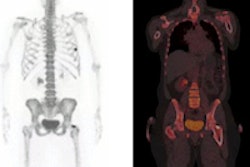
MIAMI BEACH, FL - Sodium fluoride (NaF) PET/CT detected bone metastases in patients with breast cancer more effectively than FDG, especially in cases of restaging, according to a study presented this week at the Society of Nuclear Medicine (SNM) annual meeting.
The researchers speculated that the difference in results among patients who had received therapy and those who were imaged for initial staging may be due to the functions of the two radiotracers.
"The disparity appears more pronounced and is statistically significant in the restaging population -- that is, patients who have completed or are on treatment," said study co-author Dr. Richard Wahl, director of the division of nuclear medicine/PET at Johns Hopkins University. "The difference is reflective of different mechanisms of tracer localization. The sodium fluoride represents healing, versus FDG, which represents tumor metabolism."
The lead author of the study was Dr. Muhammad Chaudhry, medical director at Tawam Molecular Imaging Center in Al Ain, Abu Dhabi, United Arab Emirates. The center is run by Johns Hopkins.
"In that practice, they use both FDG and sodium fluoride PET/CT for imaging," Wahl said during his presentation. "Clinically, there is a large prevalence of breast cancer. At this time, there are no effective screening programs, so patients present with more advanced disease."
Advanced cancer
There are also other health issues specific to younger patients who present with more-advanced cases of breast cancer.
"Clearly, bone is frequently a sign of metastatic disease," Wahl said. "Since sodium fluoride and FDG are used clinically for patients [at the center] and are paid for by the healthcare authority, it is possible to directly compare PET/CT with both FDG and sodium fluoride in the same patient ... specifically, for looking at bone metastases in women with breast cancer."
All women with a history of infiltrating ductal carcinoma of the breast underwent separate imaging with FDG-PET/CT and NaF-PET/CT between October 2011 and April 2012. In this study, 42 patients underwent 45 sequential FDG-PET/CT and NaF-PET/CT scans. On average, female patients were 51 years old, ranging in age from 30 to 77 years.
Typically, separate studies were performed within one week of each other, with 24 imaging procedures done for initial cancer staging before therapy, and 66 scans performed for restaging after treatment or surgery.
FDG vs. NaF
Among the study's patient population, for staging and restaging, FDG-PET/CT was positive for metastases in 11 cases, compared with 19 cases with NaF-PET/CT.
In a further analysis of the data, which Wahl said was somewhat limited by the small patient sample, there were three positive results with FDG-PET/CT and four with NaF-PET/CT in the staging group. Among patients who were restaged for cancer, there were eight positive FDG-PET/CT scans and 15 positive NaF-PET/CT scans, which Wahl said is "statistically significant."
"Every patient who had a positive FDG-PET/CT [also] had a positive NaF-PET/CT in the same area of the study," Wahl noted. "The difference is the significant frequency of positive [results] with NaF-PET/CT for bone metastases compared to FDG-PET."
NaF-PET/CT detected a total of 65 lesions, compared with 40 lesions for FDG-PET/CT, the researchers found.
Wahl reiterated that NaF's prowess among the restaging population may reflect "different mechanisms of tracer localization."
"I think there exists some concern in our minds that some NaF-positive lesions we detected post-therapy may represent relatively inactive tumors, at least as evidenced by FDG, given that [the tumors] were seen in the group that received treatment," he added.
Wahl and Chaudhry are currently evaluating the long-term prognostic significance of the differences between NaF and FDG, but data are not yet available.


