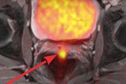
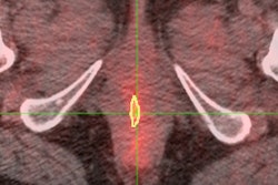
CHICAGO - PET/MRI can be comparable to PET/CT and holds "great promise" for patients with gynecological malignancies, potentially reducing scan time and radiation exposure, according to a study presented on Monday at the RSNA annual meeting.
The prospective study from the Mallinckrodt Institute of Radiology at Washington University in St. Louis found similar results in a comparison of PET/MRI and PET/CT in terms of maximum standardized uptake values (SUVmax), metabolic tumor volume, and identifying disease location.
PET/MRI also offers the ability to compare tumor boundaries between PET and various MR sequences, said lead study author Dr. Maria Thomas, PhD, chief resident in radiation oncology. "The superior soft-tissue resolution is very valuable in assessment of pelvic malignancies, and we feel this will potentially replace PET/CT for radiation treatment planning at our institution in the future," she added.
Thomas noted in her introductory remarks that PET has a strong history in cervical cancer applications and in assessing patients with gynecological malignancies.
With the addition of MRI to PET, the combined modalities have "the opportunity to visualize the integrated anatomic, metabolic, and functional imaging," she said. "Clearly, there is superior soft-tissue contrast for the pelvis and other disease sites, and the synchronous acquisition avoids differences in patient position, bladder and rectal filling, and tumor growth."
Thomas and colleagues enrolled 21 gynecological cancer patients in the study: 18 had cervical cancer, one had vulvar cancer, and two had vaginal cancer. Five patients were imaged at baseline, three were scanned during therapy, and the remaining 13 were imaged during the follow-up post-therapy period.
Patients received a single FDG injection and were first imaged using a PET/CT scanner. The participants then underwent PET/MRI exams on Siemens Healthcare's Biograph mMR, which images patents simultaneously on both modalities.
The researchers obtained SUVmax and metabolic tumor volume. Two independent reviewers scored the sites of disease in their comparison between PET/MRI and PET/CT results. They also evaluated primary tumor, lymph node, and distant metastases as positive, negative, or indeterminate.
The mean difference in SUVmax for the five patients imaged at baseline was ± 9%, according to Thomas and colleagues. The mean difference in metabolic tumor volume was ± 18%. In addition, diagnostic interpretations were similar, with no cases of positive results on one modality and negative results on the other.
Interestingly, Thomas added, the frequency of indeterminate findings was greater on PET/CT at approximately 6%, compared with approximately 2% for PET/MRI.
"In conclusion, we think that PET/MRI holds great promise for patients with gynecological malignancies," she said. "The synchronous acquisition is critical to avoiding differences in patient position, organ motion, and tumor growth."
When patients need both PET and MRI, scan time can be minimized, and radiation dose can also be reduced, the group concluded.


