Tuesday, December 3 | 10:50 a.m.-11:00 a.m. | SSG01-03 | Room E451A
German researchers from Essen University Hospital are showing positive results for PET/MRI over PET/CT for detecting and estimating the size of malignant breast lesions.PET/MRI should be the modality of choice for staging primary breast cancer, the researchers concluded. However, they found that both modalities tend to underestimate tumor size,
"Our study demonstrates the superiority of PET/MRI in detecting breast cancer lesions in estimating the size [T-stage] and the evaluation of local tumor spread, in comparison to PET/CT," said lead researcher Johannes Grüneisen. "Therefore, PET/MRI provides valuable information for decision-making on further treatment, such as surgical planning."
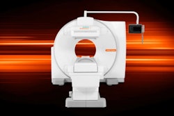
The study enrolled 43 patients with biopsy-proven invasive breast cancer. The imaging protocol included FDG-PET/CT for all patients and contrast-enhanced FDG-PET/MRI (Biograph mMR, Siemens Healthcare) using a 16-channel breast coil.
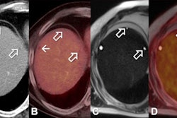
PET/CT correctly identified 46 (88%) of 52 breast cancer lesions, whereas PET/MRI correctly detected 50 (96%) of the 52 lesions. PET/MRI also correctly assessed T stage in 44 (84%) of the 52 cases; PET/CT was accurate in 31 (59%) of the 52.
The researchers found five cases in which the same lesion at T2 was falsely diagnosed as T1 by both modalities.
"Comparing the submodalities MRI and CT, MRI exhibits a higher sensitivity in assessing local breast cancer lesions due to a higher soft-tissue contrast and high spatial resolution, avoiding harmful radiation exposure," Grüneisen said. "Due to its combination with PET, PET/MR mammography provides all metabolic and morphologic information needed for local tumor staging."
In the future, Grüneisen and colleagues plan to assess the diagnostic benefit of PET/MRI versus PET/CT for whole-body staging and the detection of tumor recurrences.


