Wednesday, December 4 | 3:10 p.m. - 3:20 p.m. | SSM17-02 | Room S505AB
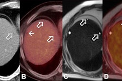
Diffusion-weighted MRI (DWI-MRI) is a viable, radiation-free alternative for evaluating lymph nodes, with findings that closely parallel those of FDG-PET/CT, researchers in India have concluded.Their study included 40 patients with a confirmed diagnosis of malignancy and significant lymph node involvement on FDG-PET/CT. These patients also underwent DWI-MRI on a 1.5-tesla system.
Evaluation of 241 lymph nodes showed a statistically significant inverse correlation between minimum apparent diffusion coefficient (ADCmin) values and maximum standardized uptake (SUVmax). There was a significant positive correlation between the visual grade of a lymph node on DWI and its PET/CT assessment, according to the researchers.
Using PET/CT as the reference standard, DWI achieved sensitivity of 90%, specificity of 90%, positive predictive value of 96%, and negative predictive value of 76%. If mediastinal lymph nodes were excluded from the analysis, DWI had sensitivity of 98%, specificity of 90%, positive predictive value of 96%, and negative predictive value of 95%.
"The study demonstrates that DWI and FDG-PET/CT parallel each other in terms of lymph node detection," said lead researcher Dr. Salil Bhargava, a senior radiology resident at Tata Memorial Hospital in Mumbai. "This will hopefully pave the way for further research in this area and allow for the development of streamlined imaging protocols for patients suffering from different cancers, all while keeping their radiation doses to a minimum."
Regarding next steps, Bhargava and colleagues plan to conduct a blinded, comparative study between DWI and PET/CT in lymphoma patients to determine if DWI alone could provide enough information for appropriate treatment decisions.


