Dear AuntMinnie Member,
Clinicians are beginning to realize that even a normal functional stress test is no guarantee that an individual is free of risk from a future heart attack. In many cases, the addition of information from an anatomical modality such as PET/CT can be useful.
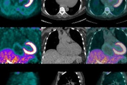
In a new study presented at this week's American Heart Association annual meeting in New Orleans, researchers demonstrated that PET/CT can find signs of coronary calcium, even in patients who have normal stress tests.
The research group from Utah installed a PET/CT scanner four years ago and immediately recognized that it was much more accurate than SPECT -- with a lower radiation dose. So the researchers decided to use the hybrid modality to study how many patients went on to have coronary events, despite having no signs of blockage on stress exams.
They found that 5% of patients classified as having severe coronary calcium went on to have a major adverse cardiac event, indicating that PET/CT could have a role in managing these individuals. Learn more by clicking here, or visit our Molecular Imaging Community at molecular.auntminnie.com.
Deep learning and elastography
The number of intriguing new applications for deep learning is growing by the day, it seems. In our Ultrasound Community this week, we're highlighting one new application: analyzing shear-wave elastography (SWE) ultrasound scans to characterize breast lesions.
Researchers from China developed a deep-learning model that learned image features on SWE scans and characterized tumors based on malignancy. They then compared the deep-learning algorithm with a semiautomated analysis method based on older technology that is typically used with computer-aided diagnosis (CADx) software.
The deep-learning model was more sensitive and specific than the CADx tool, according to the group. Find out by how much by clicking here.
While you're in the community, be sure to check out this article on how researchers developed a new ultrasound imaging mode they call H-scan, based on calculations developed by a 19th-century mathematician. These stories and more are available in our Ultrasound Community at ultrasound.auntminnie.com.
4D of ankle instability
Finally, be sure to visit our CT Community for an article on how French researchers used 4D 320-detector-row CT to assess ankle instability, in particular the subtalar joint. That story is available by clicking here, or go to ct.auntminnie.com.


