The Society of Nuclear Medicine and Molecular Imaging (SNMMI) and the American Society of Nuclear Cardiology (ASNC) have published guidelines on how to use PET imaging for the clinical quantification of blood flow.
With the aid of PET, radionuclide myocardial perfusion imaging (MPI) is designed to accurately measure total tracer activity in arterial blood and its delivery to the myocardium. By doing so, the imaging technique can evaluate regional perfusion abnormalities, dysfunction, and unusual chamber dimensions.
"Quantification of myocardial blood flow and myocardial flow reserve represents a substantial advance for diagnostic and prognostic evaluation of suspected or established coronary artery disease," the authors wrote. "These methods are at the cusp of translation to clinical practice."
The position paper, published in both the Journal of Nuclear Medicine and the Journal of Nuclear Cardiology, also noted that additional efforts are needed to standardize measures across laboratories, radiotracers, equipment, and software.
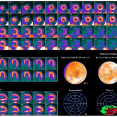
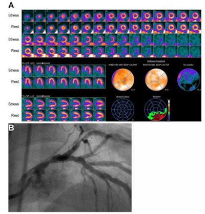 Images are from an 81-year-old man with hypertension and dyslipidemia. Myocardial perfusion imaging (A) with rubidium-82 PET showed mild, reversible perfusion abnormality in the left anterior descending coronary artery. Nearly the entire heart had severely reduced myocardial functional recovery (MFR) except for inferior and inferolateral walls, where it was moderately reduced. Coronary angiography (B) showed severe stenosis of the midportion of the left main coronary artery. Images courtesy of SNMMI and ASNC.
"Quantification of myocardial blood flow is the next great leap in nuclear cardiology, and this document summarizes the data supporting its use and offers guidance to those who are planning to make these measurements in practice," said lead author Dr. Venkatesh Murthy, PhD, vice president-elect of the SNMMI's Cardiovascular Council, in a statement.


