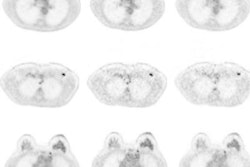
CHICAGO - PET/MRI could very well provide all the answers clinicians need to evaluate and direct treatment for patients with rectal cancer, according to a trio of studies presented on Tuesday at RSNA 2018.
Researchers from Brazil found that the hybrid modality outperformed more conventional imaging options by detecting more lesions and better assessing the spread of disease. PET/MRI also offered "tolerable scan times" and helped distinguish between high- and low-risk rectal cancer patients.
"PET/MRI for rectal cancer is feasible with a tolerable scan time and detects more lesions than conventional staging with MRI and CT," said study co-author and presenter Dr. Marcelo Queiroz from the University of São Paulo. "If we perform PET/MRI, we can combine the morphology of MRI and the metabolics of PET to identify high-risk rectal cancer patients."
Cancer staging
The purpose of the first study was to analyze the feasibility of PET/MRI for rectal cancer staging, focusing on which MR imaging protocol was most effective, total scan time, and the characteristics of the primary and metastatic tumors.
Fifty-five patients with rectal adenocarcinoma underwent whole-body PET/MRI for primary rectal cancer staging. The protocol included a dedicated pelvic MRI scan with high-resolution T2-weighted and diffusion-weighted imaging (DWI), followed by whole-body PET/MRI from head to midthigh. The scans concluded with T2-weighted imaging and DWI of the liver. Any dedicated MRI sequence was used to image the thorax.
The mean total acquisition time of PET/MRI was 64 minutes (range, 53-90 minutes). Breaking down the times further, the average PET acquisition time was 17 minutes (range, 12-25 minutes), while the MRI scan time was 52 minutes, including the dedicated pelvic and liver sequences. One patient did not complete the exam due to claustrophobia, while another patient was recalled due to movement and the resulting low image quality.
For primary lesion detection, PET and DWI were both positive in 54 (98%) of the 55 patients. Similarly, liver DWI detected 64 lesions in 12 patients, while PET identified 62 lesions in the same 12 patients.
"That is an example of DWI and PET detecting the primary tumor, so there is no clear benefit to adding DWI," Queiroz told RSNA attendees.
In addition, PET/MRI uncovered metastatic disease in 21 patients (38%). Twelve patients had disease in the liver, 10 had metastases in nonregional lymph nodes, and seven had lung metastases.
"We can say that PET/MRI for rectal cancer staging is feasible with a tolerable scan time and showing a high incidence of metastases," he concluded. "We can still optimize the protocol and shorten the acquisition time, especially by omitting isotropic T2-weighted imaging, since it does not add to the results."
Lesion detection
In the second study, the Brazilian researchers retrospectively analyzed 104 rectal cancer patients who were referred for primary staging. Their goal was to compare the detection rates of metastatic lesions using PET/MRI versus conventional staging with pelvic MRI and contrast-enhanced CT thoracic and abdominal scans.
Pelvic MRI was performed for tumor detection (T) and to detect any spread of the disease to nearby lymph nodes (N), while the thoracic and abdominal CT exams were to determine if the cancer had metastasized (M). PET/MRI was evaluated for all three measures. The researchers limited to 10 the number of lesions per organ, and follow-up and biopsy served as the reference standard.
With PET/MRI, the researchers observed 305 metastatic lesions, compared with 252 on conventional imaging. The 21% increase in detection by PET/MRI was statistically significant (p = 0.001). In nonregional lymph nodes, PET/MRI detected 73 lesions, compared with 37 lesions on conventional imaging.
Because of its higher rate of tumor detection, PET/MRI was instrumental in upgrading 15 patients (14%) with N0 disease to N1 status and changing treatment strategies.
The patient-based analysis concluded that PET/MRI confirmed positive results in 38 (40%) of 95 patients, compared with 24 (25%) with conventional imaging. PET/MRI also had fewer indeterminate lesions (10, 9%) than conventional imaging (30, 31%).
"In conclusion, we can say that for rectal cancer staging, PET/MRI represents a higher detection rate for metastatic lesions and clarifies the etiology of indeterminate lesions," Queiroz said. "This is especially relevant for this group of patients because PET/MRI may be preferable for staging because of these high detection rates."
High-risk patients
In the final study, Queiroz and colleagues looked at PET/MRI's diagnostic performance based on certain PET and MRI parameters to identify high-risk rectal cancer patients. The task is particularly important because the clinical management of this patient population depends on TNM stage. Any additional high-risk feature greatly influences the patient's treatment plan, Queiroz said.
"What we know so far about PET imaging and tumor risk is that the maximum standardized uptake value (SUVmax) is related to an advanced T stage and that the volumetric parameters of total lesion glycolysis (TLG) and metabolic tumor volume (MTV) are predictors of etiological response," he said. "But there is no association with disease-free or overall survival."
Among the 55 subjects in this study, 23 had T3 rectal cancer, 21 had T4 disease, and 10 had T2 disease.
The researchers found that TLG and MTV were significantly superior to SUVmax and SUVmean in distinguishing between low- and high-risk patients.
| PET parameters for differentiating high- and low-risk rectal cancer patients | |||
| High-risk patients | Low-risk patients | p-value | |
| MTV | 48.9 | 18.4 | 0.006* |
| TLG | 629.4 | 190.4 | 0.008* |
| SUVmax | 23.4 | 19.2 | 0.306** |
| SUVmean | 11.7 | 9.9 | 0.285** |
**No statistical significance
Patients with advanced T stage, positive regional node, and metastatic disease also had significantly higher MTV and TLG values than SUVmax and SUVmean.
"So if we perform PET/MRI, we can combine the morphology of MRI and metabolism of PET to identify these [high-risk] patients, and then direct them to the correct treatment management," Queiroz concluded.


