
A device developed by Surgi-Vision that marries MRI with cardiac radiofrequency ablation has performed better than standard x-ray-guided electrophysiologic interventions, according to a study in the August 8 issue of Circulation.
The study, by researchers at Johns Hopkins University in Baltimore, demonstrates that radiofrequency ablation (RFA) can be performed in vivo with MR-guided technology, according to Surgi-Vision, which is based in Columbia, MD.
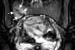
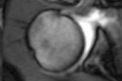
Dr. Albert Lardo and his co-authors performed RFA in the right ventricular apex of six healthy dogs. Catheters were positioned to intracardiac targets with an MR fluoroscopy sequence, and ablated tissue was imaged with T2-weighted fast spin-echo, as well as contrast-enhanced T1-weighted gradient-echo sequences.
According to the results, "ablated areas of myocardium appeared as hyperintense regions directly adjacent to the catheter tip and could be detected two minutes after RF delivery…lesion size by MR correlated well with actual postmortem lesion size and histological necrosis area" (Circulation, August 8, 2000, Vol.102, pp.698).
Some of the advantages of MR-guided RFA over traditional x-ray include the elimination of ionizing radiation exposure and a greater accuracy in therapy titration, Lardo said. In addition, MR-guided RFA could provide insight into the physiological effects of ablation, he added.
Surgi-Vision received U.S. Food and Drug Administration clearance for marketing its miniature internal coil technology in 1999.
By AuntMinnie.com staff writers
August 10, 2000
Related Reading
Surgi-Vision gets clearance for coil, April 5, 2000
GE signs deal with Surgi-Vision
Copyright © 2000 AuntMinnie.com


.fFmgij6Hin.png?auto=compress%2Cformat&fit=crop&h=100&q=70&w=100)

.fFmgij6Hin.png?auto=compress%2Cformat&fit=crop&h=167&q=70&w=250)


