After enjoying a honeymoon period, computer-aided detection (CAD) technology has begun to receive more scrutiny, especially as it moves out from mammography into other modalities.
Researchers at the department of medical imaging at Sunnybrook Health Sciences Centre in Toronto evaluated the effect of MRI CAD on radiologists' sensitivity and specificity for breast lesions recommended for biopsy after MR screening in women with a high risk of breast cancer. They presented their study findings at the 2007 RSNA meeting in Chicago.
Dr. Tal Arazi-Kleinman and colleagues took 56 consecutive BI-RADS 3 to BI-RADS 5 lesions with histopathologic correlation that had been detected in high-risk patients with MRI screening and retrospectively evaluated them with MR CAD. The pool of lesions included the following:
- 9 invasive cancers
- 13 ductal carcinoma in situ (DCIS)
- 34 benign lesions
Two breast imaging specialists performed the CAD evaluations with lesion enhancement at thresholds of 50%, 80%, and 100%, as well as with delayed enhancement. Histopathology characteristics were blinded. The lesions were rated as malignant or benign in relation to background parenchymal enhancement according to threshold, delayed enhancement only, and in combination. The group determined sensitivities and negative predictive values (NPV) for the final CAD assessments versus pathology for the invasive and DCIS lesions separately.
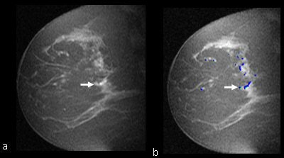 |
| MR images, without and with CAD, applied in a 50-year-old patient with a strong family history of breast cancer with a left breast MRI-detected benign lesion, diagnosed at MRI-guided vacuum assisted biopsy. (a) Sagittal T1-weighted 2D spoiled gradient recalled (SPGR) fat-suppressed (150/4.2; flip angle, 50°; matrix, 256 x 128; field-of-view 20 cm; slice thickness, 3 mm) image shows clumped linear enhancement (arrow) in the central left breast. (b) CAD assessment of the lesion demonstrated enhancement at all the threshold levels. At 80% threshold (arrow) CAD color mapping of the delayed enhancement pattern shows predominantly blue with some green, indicating a persistent delayed enhancement pattern that did not differ from the remaining background enhancement. This was therefore assessed as benign based on CAD. All images courtesy of Dr. Tal Arazi-Kleinman, Sunnybrook Health Sciences Centre. |
Arazi-Kleinman and colleagues found that discriminatory threshold levels for lesion enhancement were 50% for all cancers, DCIS, and all benign lesions, and 100% for all invasive cancers. The combined use of threshold and enhancement pattern data for CAD assessment was best, with 73% sensitivity, 56% specificity, and 76% NPV for all cancers.
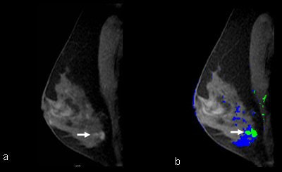 |
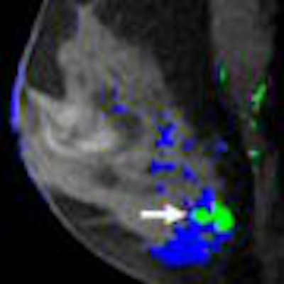
| MR images, with and without CAD, applied in a 49-year-old BRCA1 patient with a left breast MRI-detected invasive ductal carcinoma, diagnosed at MRI-guided wire localization and surgical excisional biopsy. (a) Sagittal T1-weighted 2D SPGR fat-suppressed (150/4.2; flip angle, 50°; matrix, 256 x 128; field-of-view 18 cm; slice thickness, 3 mm) image shows a heterogeneous enhancing lobulated mass (arrow) in the central lower left breast. (b) CAD assessment of the lesion demonstrated enhancement at the all the threshold levels. At 80% threshold CAD color mapping of the delayed enhancement pattern shows predominantly green with a dot of red that differs from the mainly blue background. This was therefore assessed as malignant based on CAD. |
But sensitivities and NPV were better for invasive cancers only (100% and 100%, respectively), compared to DCIS only (54% and 76%, respectively). The researchers found that radiologists' MRI interpretation was more sensitive than CAD but less specific for cancer detection in a high-risk population.
Current breast MR CAD cannot improve radiologist accuracy in distinguishing malignant from benign MRI screen-detected lesions because of its poor DCIS detection sensitivity, they concluded.
By Kate Madden Yee
AuntMinnie.com staff writer
February 18, 2008
Related Reading
Preop MRI can improve breast cancer surgical outcomes, study says, December 27, 2007
Breast MRI comparable to mammography for detecting later cancers, November 25, 2007
Breast MR centers must look beyond imaging and offer universal care, November 2, 2007
MRI beats US for breast screening of at-risk women, but yields more biopsies, August 6, 2007
Breast MRI helps guide surgical treatment, May 22, 2007
Copyright © 2008 AuntMinnie.com




.fFmgij6Hin.png?auto=compress%2Cformat&fit=crop&h=100&q=70&w=100)



.fFmgij6Hin.png?auto=compress%2Cformat&fit=crop&h=167&q=70&w=250)

