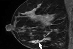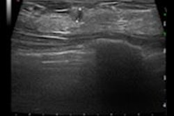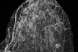
Elevated levels of background parenchymal enhancement (BPE) on breast MRI increase a woman's risk of developing invasive breast cancer -- even more than breast tissue density does, according to a study published online January 9 in the Journal of Clinical Oncology.
Furthermore, the combination of elevated BPE and dense breast tissue increases a woman's overall risk for breast cancer more than either factor on its own, wrote a team led by Dr. Vignesh Arasu of Kaiser Permanente Medical Center in Vallejo, CA.
"Among women undergoing screening or diagnostic breast MRI, elevated levels of BPE significantly increased risk of developing primary invasive breast cancer," the group wrote. "BPE had a stronger association with breast cancer risk than breast density in this population. Moreover, BPE was independent of breast density in risk prediction, and the combination of BPE and breast density increased the overall risk for breast cancer more than either factor alone."
Arasu and colleagues evaluated the association of breast MRI background parenchymal enhancement and breast density with subsequent breast cancer risk through a study that included 4,247 women undergoing breast MRI in facilities participating in the Breast Cancer Surveillance Consortium (BCSC) from 2005 to 2015.
The women's BPE was assessed at the time of interpretation of the breast MRI exam. The team used mammography results performed within five years of the MRI scans to assess the women's breast tissue density and risk factors. The group estimated hazard ratios for breast cancer using a BCSC model.
Among the 4,247 women included, 176 developed breast cancer over a median follow-up period of 2.8 years. Of these cancers, 129 were invasive (73%) and 47 were ductal carcinoma in situ (27%).
The team found that increasing BPE levels were associated with significantly increased cancer risk, as was tissue density.
| Association between BPE and breast tissue density and cancer risk | |||
| Women with cancer (n = 176) | Women without cancer (n = 4,071) | Hazard ratio | |
| BPE in 2 categories | |||
| Minimal | 20% | 34% | -- |
| Mild, moderate, or marked | 80% | 66% | 2.28 |
| Breast density | |||
| Fatty | 4% | 5% | 1.19 |
| Scattered fibroglandular | 23% | 30% | -- |
| Heterogeneously dense | 41% | 43% | 1.29 |
| Extremely dense | 31% | 22% | 1.54 |
| Density and BPE | |||
| Low density and minimal BPE | 10% | 16% | -- |
| High density and minimal BPE | 10% | 18% | 1.25 |
| Low density and mild, moderate, or marked BPE | 18% | 19% | 2.30 |
| High density and mild, moderate, or marked BPE | 62% | 47% | 2.61 |
| BCSC 5-year risk and BPE | |||
| BCSC risk < 1.67% and minimal BPE | 6% | 17% | -- |
| BCSC risk ≥ 1.67% and minimal BPE | 12% | 17% | 1.79 |
| BCSC risk < 1.67% and mild, moderate, or marked BPE | 33% | 35% | 2.91 |
| BCSC risk ≥ 1.67% and mild, moderate, or marked BPE | 49% | 32% | 4.03 |
"Women with a higher BPE and a high BCSC five-year risk score of 1.67% or greater had a fourfold increased risk [of breast cancer] compared with women with minimal BPE and a low risk score," Arasu and colleagues noted.
BPE offers a promising way to predict future breast cancer risk, the team concluded.
"BPE should be considered for incorporation into risk prediction models for women undergoing MRI," the group wrote.


