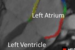
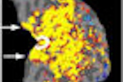
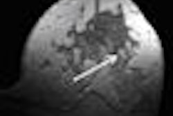
University of Utah researchers have found that delayed-enhancement MRI (DE-MRI) holds promise for predicting treatment outcomes and measuring disease progression for patients with atrial fibrillation (AF).
The study, published in the April 7 issue of the journal Circulation, assessed DE-MRI in a protocol developed to detect fibrosis in atrial fibrillation patients before they underwent radiofrequency ablation.
The researchers from the Salt Lake City university developed a protocol to create 3D MR images of the left atrium before radiofrequency ablation, which were processed and analyzed with custom software tools and computer algorithms to calculate the extent of left atrium wall injury.
Six months after the procedure, the researchers found that only 14% had minimal fibrosis and suffered atrial fibrillation recurrence, compared with 75% recurrence for the group that had extensive scar tissue damage.
According to the study, the results "indicate that DE-MRI provides a noninvasive means of assessing left atrial myocardial tissue in patients suffering from atrial fibrillation, and that those who do have tissue damage may be at greater risk of suffering atrial fibrillation recurrence after treatment with radiofrequency ablation."
Related Reading
Atrial fibrillation tied to cognitive impairment in patients without stroke, September 26, 2008
MRI evaluation of chest pain cuts acute coronary syndrome, August 15, 2008
Screening detects additional cases of atrial fibrillation, August 3, 2007
Obesity increases radiation dose delivered during atrial fibrillation ablation, July 20, 2007
Copyright © 2009 AuntMinnie.com


