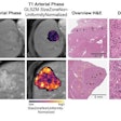
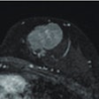
Pediatric neuroscientists at Oregon Health & Science University's Doernbecher Children's Hospital are having early success in viewing tiny white-matter injuries with 12-tesla MRI in premature fetal sheep, suggesting the modality could potentially be used to image the brains of preterm infants, according to a study published in the Annals of Neurology.
The ability to accurately identify these lesions could prevent delays in therapy, as well as allow physicians to inform families sooner of the potential for complications.
Principal investigator Dr. Stephen Back, PhD, associate professor of pediatrics and neurology in the Papé Family Pediatric Research Institute, and colleagues noted that white-matter injury is the most common cause of chronic neurologic disability in children with cerebral palsy.
Prior to this study, the ability to develop treatments for white-matter injury in preterm infants has been hampered by clinicians' inability to see these microscopic injuries. One tiny lesion can have a tremendous impact on the patient's ability to walk and learn.
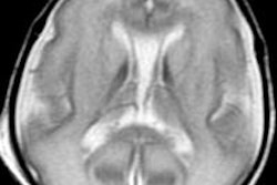
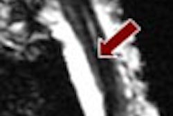
However, using 12-tesla MRI, Back and colleagues identified tiny brain lesions in preterm fetal sheep with characteristics previously unseen and unreported using a standard 3-tesla MRI.
The findings support the potential of using high-field MRI for early identification and improved diagnosis and prognosis of white-matter injury in preterm infants, Back said. The "preclinical animal model provides unique experimental access to questions directed at the cause of these lesions, as well as the optimal field strength and modality to resolve evolving lesions using MRI," he added.
Future studies are needed to determine the clinical-translational utility of high-field MRI, he said.


.fFmgij6Hin.png?auto=compress%2Cformat&fit=crop&h=100&q=70&w=100)


.fFmgij6Hin.png?auto=compress%2Cformat&fit=crop&h=167&q=70&w=250)


