Sunday, November 25 | 11:55 a.m.-12:05 p.m. | SSA16-08 | Room N228
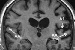
Functional MRI could provide a better understanding of how the human brain deteriorates and attention function declines as a person ages, according to a Chinese study to be presented on Sunday.This study at the Drum Tower Hospital of Nanjing University included one group of 18 younger individuals and a second group of 22 older adults who received echo-planar functional MRI exams.
Researchers analyzed the two subtypes of frontotemporal lobar degeneration (FTLD) -- behavioral-variant frontotemporal dementia (bvFTD) and primary progressive aphasia (PPA) -- and evidence of Alzheimer's disease to identify patterns of cortical atrophy in the brain.
A high-resolution 3D T1-weighted turbo field-echo (TFE) sequence was used to calculate the volume of gray matter, and connectivity maps were compared between the two groups.
In the analysis, functional MR images revealed a significant decrease in attention network connectivity in older subjects compared with the younger group.
"Among them, behavioral-variant frontotemporal dementia had asymmetric right frontal and temporal lobe atrophy, and related to characteristic personality changes," said lead study author Dr. Hongying Zhang. "Otherwise, the asymmetric atrophy in the left temporo-occipital lobe explained the aphasis of patients with primary progressive aphasia. Therefore, the MRI might be a useful marker for the pathological changes in the brain with frontotemporal lobar degeneration."
Zhang and colleagues are continuing their research and plan to use diffusion-tensor MRI and PET/CT to collect additional data.


.fFmgij6Hin.png?auto=compress%2Cformat&fit=crop&h=100&q=70&w=100)



.fFmgij6Hin.png?auto=compress%2Cformat&fit=crop&h=167&q=70&w=250)
