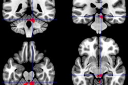
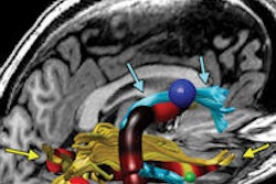
A study using functional MRI (fMRI) has found that patients with chronic fatigue syndrome may have reduced responses in a brain region associated with fatigue.
Compared with healthy controls, patients with chronic fatigue syndrome (CFS) had less activation of the basal ganglia, and the reduced activation was also linked with the severity of fatigue symptoms. The findings suggest that CFS is associated with changes in brain circuits that regulate motor activity and motivation (PLOS One, May 23, 2014).
The researchers compared 18 patients with chronic fatigue syndrome with 41 healthy volunteers. For the brain imaging portion of the study, participants were told they would win a dollar if they correctly guessed whether a preselected card was red or black. After they made a guess, the color of the card was revealed; at that moment, the researchers measured blood flow to the basal ganglia.
The key measurement was the difference in activity between a win or a loss. Participants' scores on a survey gauging their levels of fatigue were tied to the difference in basal ganglia activity between winning and losing. Those with the most fatigue had the smallest changes, especially in the right caudate and the right globus pallidus.
Previous studies have suggested a link between viral infection and CFS, said lead author Dr. Andrew Miller from Emory University School of Medicine. The current study supports the idea that an immune response to viruses could be associated with fatigue by causing inflammation in the brain.
Ongoing studies at Emory are further investigating the impact of inflammation on the basal ganglia.


