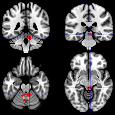
Functional MRI (fMRI) has helped uncover significant differences in brain activity between individuals with Gulf War illness and chronic fatigue syndrome, despite having similar symptoms of malaise and pain, in two studies presented Wednesday at the Society for Neuroscience annual meeting in Chicago.
Researchers from Georgetown University Medical Center confirmed opposite reactions and activity in certain brain regions of subjects with either Gulf War illness or chronic fatigue syndrome. This information could provide a foundation to effectively diagnose the conditions and develop appropriate treatments.
"This new work further emphasizes that chronic fatigue syndrome and Gulf War Illness are two very real, and very distinct, diseases of the brain," said researcher Dr. James Baraniuk in a statement.
Very similar symptoms
It is estimated that approximately 233,000 veterans of the Persian Gulf War in 1990-1991 developed Gulf War illness after exposure to toxic nerve agents, pesticides, and other neurotoxins. On the other hand, the etiology of chronic fatigue syndrome remains unknown. Still, the two conditions have very similar symptoms, including sleep and cognitive dysfunction, negative emotions, gastrointestinal problems, pain, and fatigue. All of the symptoms often worsen after physical exercise.
To further investigate how these conditions affect brain function, the researchers conducted two studies involving neuronal brain activity using fMRI and a series of cognitive tests before and after exercise.
In the first study, the researchers divided subjects into three groups: 80 patients with Gulf War illness, 38 people with chronic fatigue syndrome, and 23 healthy controls. All 141 participants underwent 3-tesla fMRI scans and continuous N-back memory tasks, followed by a bicycle stress test and a second stress test 24 hours later, to see how specific brain areas are affected by the disorders.
"As Gulf War illness and chronic fatigue syndrome are both characterized by postexertional malaise, all three groups completed fMRI scans before and after two exercise stress tests," the researchers explained in their abstract.
Exercise effects
Before exercise, fMRI showed significantly greater activation in the right angular gyrus brain region for patients with Gulf War illness compared with the other two groups. After exercise, patients with Gulf War illness had significantly less activation in the cerebellar vermis and right intraparietal sulcus than their counterparts.
Conversely, fMRI revealed significantly greater activation in the left rolandic operculum among chronic fatigue syndrome subjects compared with the other two groups and greater activation than Gulf War illness cases in both the periaqueductal gray and right insula. The researchers found no significant changes in brain region activation before or after exercise among the healthy control subjects.
"In short, peri-insular neural activity differentiates chronic fatigue syndrome from Gulf War illness and controls, whereas medial cerebellar activity differentiates Gulf War illness from chronic fatigue syndrome and controls," they noted. "These findings suggest qualitatively different neural substrates of postexertional malaise associated with cognition in Gulf War illness and chronic fatigue syndrome."
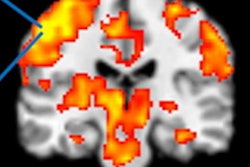
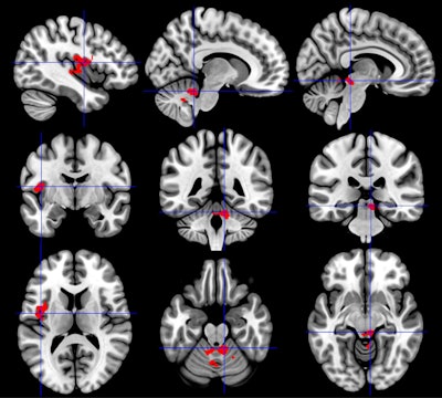 The insula region of the brain (right column) has greater activation in individuals with chronic fatigue syndrome than in healthy controls or those with Gulf War illness. By comparison, the cerebellum (middle column) has less activation in Gulf War illness than healthy controls or those with chronic fatigue syndrome. In addition, the brainstem showed less activation (left column) in Gulf War illness and greater activation in chronic fatigue syndrome. All the differences were observed after exercise. Images courtesy of Stuart Washington, PhD.
In the second study, the researchers compared brain region activation based on blood oxygenation level dependent (BOLD) contrast among 79 veterans with Gulf War illness, 36 people with chronic fatigue syndrome, and 31 healthy controls. The goal was to explore nine brainstem regions of interest and four media structures, all of which are linked to memory performance, to illustrate the degree to which these conditions have differing effects.
Again, all subjects underwent 3-tesla fMRI scans before and after two exercise stress tests. Cognitive evaluations were done through 2-back working memory tasks. The researchers targeted nine brainstem regions of interest and four media structures, all of which are linked to the ability to determine threats, arousal, modulation of chronic pain, sleep, and other neurobehavioral functions.
Before exercise, fMRI showed significantly decreased BOLD activity among chronic fatigue syndrome subjects compared with healthy controls in the pedunculopontine nucleus, which is associated with learning, reward, and voluntary arm and leg movement. All other brainstem regions showed no significant difference in activity among the three groups. After exercise, Gulf War illness patients had significantly reduced BOLD activity compared with chronic fatigue syndrome subjects in eight brain regions, while no significant difference was found in healthy controls.
The results in this study support other research conducted postmortem in veterans with post-traumatic stress disorder and suggest the brainstem in these veterans may have physical abnormalities, such as a loss of neurons.
"The midbrain is affected by the exercise and cognitive challenges, but chronic fatigue syndrome and Gulf War illness react in opposite ways, showing that they are related but distinctly different disorders," said Haris Pepermintwala, a research student at Georgetown University, who presented the results at the meeting.


