Improved signal-to-noise ratio and anatomical detail are making MRI a valuable tool for evaluating fetuses suspected of having congenital anomalies.
That conclusion comes from researchers at the Mayo Clinic in Jacksonville, FL, who presented their work at this week's American Roentgen Ray Society (ARRS) meeting in Toronto.
Lead author Dr. Kathleen Carey said 3-tesla MRI could provide better visualization of subcutaneous fat and osseous structures, including the hands and feet of the developing fetus, but the benefits must be weighed against potential risks to the fetus from the higher field strength.
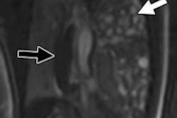
Carey presented a series of fetal MRI cases performed on a 1.5-tesla scanner (Signa HDxt, GE Healthcare), discussing the findings, diagnosis, and effect on patient management, along with other aspects of MRI.
At the Mayo Clinic, MRI sequences are monitored in real-time by a radiologist at the scanner and manipulated to determine optimal axial, coronal, and sagittal imaging planes during the scan. Images are typically obtained after 18 to 20 weeks of gestation, and no gadolinium is used.
It is important to understand the indications and limitations of fetal MRI at 1.5 tesla and higher field strengths, as fetal imaging continues to progress toward 3-tesla scanning, the group concluded.


.fFmgij6Hin.png?auto=compress%2Cformat&fit=crop&h=100&q=70&w=100)


.fFmgij6Hin.png?auto=compress%2Cformat&fit=crop&h=167&q=70&w=250)
