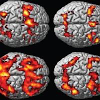
A new study published online April 28 in Radiology indicates that women have a more difficult time recovering from concussions than men, based on brain activation patterns seen on functional MRI (fMRI) during working memory tasks. The results hint that more aggressive treatment could be warranted after head injuries in women.
Six weeks after mild traumatic brain injury (MTBI), the brains of men had returned to normal activation patterns similar to those of a control group, while women showed less brain activation, or persistent hypoactivation, indicating continued problems with working memory.
Senior author Dr. Chi-Jen Chen and colleagues from Taipei Medical University Shuang-Ho Hospital said their work may be the first published evidence of differences in working memory functional activity between the two genders in patients with MTBI.
In addition, functional MRI "has the potential to not only provide useful objective diagnostic information associated with working memory functional sequelae, but also to provide a sensitive measurement with which to monitor disease progression and treatment," they wrote (Radiology, April 28, 2015).
MTBI incidents
While most patients with concussions fully recover within three months, as many as 10% to 15% will have lingering health issues, such as headaches, interrupted sleep, loss of balance, memory and other cognitive impairments, and fatigue.
Chen and colleagues noted that working memory impairment is a common problem after a concussion and can adversely affect daily life. No previous research had been done to determine if there is a difference in recovery time between men and women, they added.
The current study included 15 consecutive men and 15 consecutive women with MTBI who presented to the emergency department for a number of incidents, including motor vehicle collisions, falls, sports injuries, and assault. In addition, 30 healthy control subjects (also 15 consecutive men and 15 consecutive women) were recruited for the study.
Potential subjects were excluded for a number of reasons, including prior traumatic brain injury, any other neurologic disease, current or past history of psychiatric illness, current use of psychoactive medications, and any contraindications to MRI.
Two fMRI scans were performed on a 3-tesla scanner (Discovery MR750, GE Healthcare) with an eight-channel receiver-only head coil. The first exam was performed within one month of the injury, and a follow-up scan was conducted six weeks after the initial MRI.
Neuropsychological assessments were given to all 60 subjects, including a short-term memory test known as a digit span, which measures how many numbers a person can remember in sequence. The researchers also used a continuous performance test (CPT) to measure sustained and selective attention and impulsivity. The digit span and CPT assessments were performed before the two working memory task fMRI studies.
Gender comparisons
There was no significant difference between male patients with MTBI and male control subjects in terms of digit span or CPT results, the authors wrote.
Among the female participants, however, those with MTBI had lower digit span scores than the control subjects. There was no significant difference in CPT results between the two groups of women.
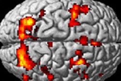
Initial fMRI scans of the MTBI patients showed increased activation in working memory brain circuits in the men and decreased activation in the women, compared with the controls.
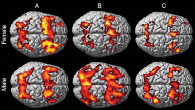 The images show increased activation in bilateral frontal and parietal regions, which is consistent with activation of working memory circuitry in each group. In control subjects (A), visual comparison of working memory activation patterns shows more activation in women than in men, especially in the frontal region. There is substantially less activation (hypoactivation) in female patients with MTBI (B) than in female control subjects, as well as substantially more activation (hyperactivation) in male patients with MTBI than in male control subjects. At the six-week follow-up study (C), female patients with MTBI showed persistent hypoactivation patterns, whereas male MTBI patients showed regression of hyperactivation and activation levels similar to those of male control subjects. Image courtesy of Radiology.
Most significantly, at the six-week follow-up fMRI scan, female patients with MTBI showed persistent hypoactivation, which suggests ongoing working memory problems. In contrast, the hyperactivation in the male MTBI patients had regressed and they had returned to a more normal level of activation, similar to what was seen in the control subjects.
Hypoactivation can be caused by more-severe brain damage, which does not allow another area of the brain to compensate for the loss of function.
"The results suggest that women may have worse working memory outcomes, and functional MR imaging showed sex differences in postinjury changes," the authors wrote. "These results may change the future diagnostic workup in patients [with] MTBI and lead to separate management strategies for patients of different sexes."
Because the results are preliminary, the group recommends additional research with a larger patient sample and a host of imaging exams, including diffusion-tensor MRI, to evaluate the brain's structural integrity, functional connectivity, and cerebrovascular reactivity to better understand the gender differences.
If it is confirmed that women do have a more difficult time recovering from concussions, "more aggressive management should be initiated once MTBI is diagnosed in female patients," the researchers wrote.


.fFmgij6Hin.png?auto=compress%2Cformat&fit=crop&h=100&q=70&w=100)

.fFmgij6Hin.png?auto=compress%2Cformat&fit=crop&h=167&q=70&w=250)


