Diffusion-tensor MR images (DTI-MRI) hold the key to predicting which concussed patients are most likely to fully recover from their head injuries as soon as a year's time, according to a study published online June 9 in the American Journal of Neuroradiology.
By also seeing how the brain compensates for concussion damage, caregivers can more quickly begin the appropriate therapy to hasten favorable outcomes, according to researchers from Albert Einstein College of Medicine and Montefiore Health System in New York City.
"While we still lack effective treatments, we now have a better understanding of the neurological mechanisms that underlie a favorable response to concussion, which opens a new window on how to look at therapies and to measure their effectiveness," said study leader Dr. Michael Lipton, PhD, professor of radiology, psychiatry and behavioral sciences, and neuroscience at Albert Einstein College and director of MRI services at Montefiore, in a prepared statement.
Physicians have had no reliable way to differentiate in advance which patients might have lingering, long-term adverse effects from those who would have a complete recovery, added lead author Dr. Sara Strauss, a resident in the department of radiology at Montefiore. As many as 15% to 30% of patients experience concussion symptoms indefinitely.
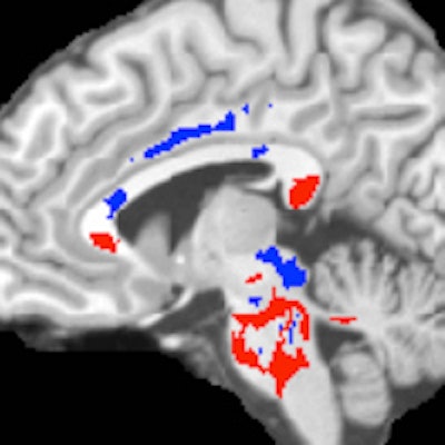
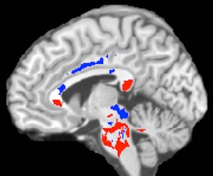 DTI-MRI of a concussion patient's brain. Areas of low fractional anisotropy (red) indicate injured white matter, while high fractional anisotropy areas (blue) suggest more efficient white-matter connections compensating for concussion damage. A large amount of high fractional anisotropy can predict recovery from a concussion. Image courtesy of Albert Einstein College of Medicine.
The current paper follows a 2009 study by Lipton et al in which the researchers showed DTI-MRI's ability to detect concussion-related damage to nerve fibers in the brain. DTI-MRI tracks the movement of water molecules along the nerve fibers, known as axons, allowing researchers to measure the flow via fractional anisotropy. Low fractional anisotropy values indicate disrupted flow and structural brain damage.
In the current study, Lipton and colleagues performed DTI scans on 39 patients diagnosed with mild traumatic brain injury within 16 days of their concussions, as well as 40 healthy control subjects. Study participants were evaluated for cognitive function, postconcussion symptoms, and health-related quality of life. One year later, 26 concussed patients returned for follow-up assessments (Am J Neuroradiol, June 9, 2016).
When the concussed patients were compared with the healthy controls, DTI-MRI showed areas of abnormally low fractional anisotropy that correlate with nerve fiber damage and cognitive impairment in concussed patients.
The images also showed other brain areas with abnormally high fractional anisotropy, which may suggest that the brain was responding to the injury by remyelinating, or repairing, injured tissue. Most importantly, having a greater volume of white-matter areas with higher fractional anisotropy correlated with better outcomes and recovery times for concussed patients.
Conversely, the areas of low fractional anisotropy, indicating white-matter damage, were not useful in predicting recovery from concussion one year later.
The findings suggest that concussion therapy may be more effective if it targets the brain's own ability to compensate or repair white-matter damage, rather than trying to minimize the damage of the concussions, Lipton said. Further studies are needed to validate these results, he added.


