Two key characteristics on MRI scans of rectal cancer patients can help predict long-term survival and local recurrence of the disease, according to a study by researchers from China and published in the August issue of the American Journal of Roentgenology.
By using MRI to gauge extramural venous invasion and circumferential resection margin, the researchers found prognostic indicators that can predict which patients have a higher risk for recurrence of rectal cancer.
"Estimating the presence of risk factors with preoperative MRI can help predict prognosis in patients with rectal cancer and determine whether patients need to receive neoadjuvant chemotherapy," wrote lead author Xiao-Xuan Jia and colleagues from Peking University People's Hospital in Beijing (AJR, August 2018, Vol. 211:2, pp. 327-334).
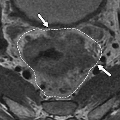
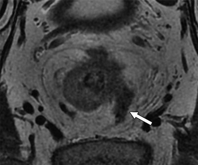
Extramural venous invasion status is a key component in the evaluation of a tumor surrounded by muscle or containing red blood cells. Circumferential resection margin is used to determine the risk of local recurrence in an excised tumor.
The retrospective study included 185 patients (mean age of 63.3 ± 11.77 years) with confirmed rectal cancer between January 2006 and September 2014 but who had not yet received neoadjuvant chemotherapy. Rectal MRI scans were performed on a 3-tesla system with an eight-channel torso array coil prior to surgery.
 Above: MR image from a 55-year-old man with rectal cancer. Arrow indicates extramural venous invasion on T2-weighted image. Below: Rectal cancer in a 70-year-old man is evident (arrows and dotted line) using circumferential resection margin involvement on T2-weighted image. Images courtesy of AJR.
Subjects were divided into a high-risk group (120 patients, 65%), with risk determined by factors such as a T3-stage tumor with a depth of more than 5 mm or T4-stage tumor, and a low-risk group (65 patients, 35%) with factors such as T1-, T2-, or T3-stage cancer with a depth of 5 mm.
Circumferential resection margin on MRI was deemed to be a significant indicator of overall survival, disease-free survival, and local recurrence. In fact, rectal cancer patients were five times more likely to survive over a median follow-up time of 42 months based on the hazard ratio. Extramural venous invasion was significantly influential regarding disease-free survival but not so with overall survival and local recurrence of the disease.
| Key MRI-based factors for rectal cancer outcome | ||||
| Circumferential resection margin | Extramural venous invasion | |||
| Hazard ratio | p-value | Hazard ratio | p-value | |
| Overall survival | 4.78 | 0.02* | 2.48 | 0.16** |
| Disease-free survival | 2.44 | 0.01* | 2.46 | < 0.01* |
| Local recurrence | 3.92 | 0.04* | 1.60 | 0.45** |
**Not statistically significant
To gauge the efficacy of postoperative chemoradiotherapy, the researchers tallied 24 patients (37%) in the low-risk group who underwent treatment but found no significant difference in the three-year overall survival, disease-free survival, or local recurrence compared with patients who did not undergo therapy.
The researchers cited several limitations of the study, including patient enrollment period that extended over several years, during which the "quality of surgery and MRI scanning techniques has changed substantially over time," they noted.


