Researchers from Rice University and MD Anderson Cancer Center are developing a way to load iron inside nanoparticles to create MRI contrast agents that potentially could outperform gadolinium chelates, according to a study published online August 8 in ACS Nano. The particles are based on nanomatryoshkas, concentric layered nanoparticles that draw their name from Russian nesting dolls.
The latest work is part of ongoing efforts to create light-activated nanoparticles for therapeutic and diagnostic purposes. The theranostic particles could allow clinicians to diagnose and treat cancer.
"Iron chelates aren't new," said lead researcher Naomi Halas, PhD, director of Rice's Laboratory of Nanophotonics, in a statement from the university. "It's widely believed they are wholly impractical for T1 contrast, but this study is a perfect illustration of how differently things can behave when you engineer at the nanoscale."
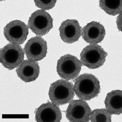
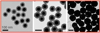 From left to right, images from a transmission electron microscope show how nanomatryoshkas are created, beginning with gold cores, progressing through the addition of a silica layer, and followed by a shell of gold. Image courtesy of Luke Henderson/Rice University.
Gadolinium chelates have been part of contrast-enhanced MRI scans since the late 1980s. More recently, however, gadolinium-based contrast agents (GBCAs) have been the focus of considerable research, following the discovery of residual gadolinium traces in brain tissue and bone long after GBCA-enhanced MRI scans.
"We were hearing more calls from the medical community for alternatives to gadolinium, and we decided to try iron chelates and see if we got the same sort of enhancement," added Luke Henderson, a Rice graduate student and lead author of the current study. "If clinicians could visualize the [iron] particles through some sort of imaging, therapy could be faster and more effective."


