Three new techniques for MRI-guided high-intensity focused ultrasound (HIFU) can more exactly target an area in the brain linked to Parkinson's disease and essential tremor and allow for more precise treatment of these conditions, according to a review published June 14 in Brain.
The techniques could translate into better patient outcomes without surgery, said lead author Dr. Bhavya Shah of the University of Texas Southwestern in Dallas in a statement released by the university.
"The benefit for patients is that we will be better able to target the brain structures that we want," he said. "And because we're not hitting the wrong target, we'll have fewer adverse effects."
Imprecise targeting of diseased brain tissue in Parkinson's or essential tremor patients can result in complications such as problems walking or slurred speech -- which tend to be temporary but can be permanent in 15% to 20% of cases, Shah and colleagues noted.
The basic protocol of MRI-guided HIFU is already approved by the U.S. Food and Drug Administration (FDA) for essential tremor and tremor-dominant Parkinson's disease, according to the university; it is based on standard MRI sequences that don't clearly identify the ventral intermediate nucleus, dentatorubrothalamic tract, and other deep brain nuclei, the team wrote.
In their review, the researchers outlined three new MRI techniques that improve MRI-guided HIFU's performance:
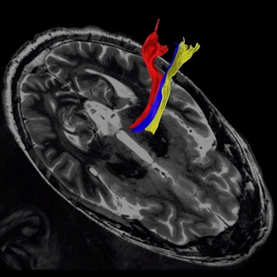
- Diffusion tractography, an FDA-approved method that creates 3D white-matter maps from diffusion-weighted imaging (DWI) -- which is an MRI sequence sensitive to water movement within tissues. "DWI can serve as an indirect measurement of anisotropy and structural organization of white matter," the group wrote.
- Quantitative susceptibility mapping (QSM), which creates contrast in MRI images by identifying distortions in the magnetic field caused by iron or blood. "Recent studies have shown that QSM can delineate thalamic nuclei with superior detail when compared to other MRI sequences," Shah and colleagues wrote.
- Fast gray-matter acquisition T1 inversion recovery, which creates a "photo negative" of the MRI image, with white brain matter appearing dark and gray matter appearing white to show greater detail.
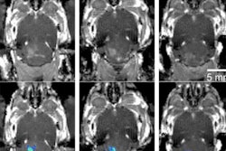
 Diffusion tractography uses the movement of water molecules to identify tracts that connect different parts of the brain. It can be used to pinpoint the part of the thalamus to treat with focused ultrasound. Image courtesy of the University of Texas Southwestern Medical Center.
These techniques show promise in identifying diseased tissue caused by Parkinson's or essential tremor, the group wrote. In fact, Shah and colleagues plan to participate in a multicenter clinical trial with collaborators at the Mayo Clinic in Rochester, MN, testing the diffusion tractography method in patients, according to the university statement.
"These advancements should lead to improved clinical efficacy, a reduction of adverse effects, and a new era of noninvasive neural modulation based on the principles of circuit biology," the authors concluded.


