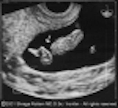
First-trimester sonograms could give obstetricians a valuable jump in detecting fetal malformations and genetic problems in at-risk women, according to an international project that is tracking the earliest gestational age at which hundreds of fetal anomalies can be detected.
So far, researchers have determined that 383 types of fetal problems can be detected as early as between 8 and 16 weeks. The knowledge has prompted them to conclude that at-risk women should receive a comprehensive sonogram sometime in the first trimester, according to a study presented at the 2001 American Institute of Ultrasound in Medicine meeting in Orlando.
The finding represents the preliminary fruits of the International Registry of Fetal Anomalies (IRONFAN), a multinational collaboration of 88 fetal imaging centers that is collating a database of sonographically detected anomalies, as well as related information concerning the management and outcome of each case.
"We are trying to unveil the natural history of fetal malformations using ultrasound. Like other institutions mapping the genome, we’re mapping the fetus with ultrasound," said IRONFAN director Dr. Shraga Rottem of Columbia University’s Audubon Biomedical Research Center in New York City.
Ultimately, this database of first-trimester problems, which so far encompasses more than 2,000 cases of fetal malformations detected at 8 to 16 weeks, could initiate a fundamental shift in how at-risk pregnancies are monitored, Rottem explained.
Rather than scanning all women when they’re between 18 to 20 weeks into the pregnancy, women with a history of fetal malformations or who are at risk of fetal problems should receive a sonogram as soon as their fetus’ potential anomaly can be detected -- which can be as early as 8 weeks.
As Rottem puts it, sonograms shouldn’t be administered according to HMO-based reimbursement schemes. Instead, they should be administered in a time-oriented manner, meaning they should target anomalies as they appear during fetal development. An anomaly that first appears at 10 weeks should be scanned at 10 weeks.
"Usually, we screen for diseases when we know it’ll show up on a test. If it’s a neonatal disease, we screen when the child is six months old, if it’s a disease that strikes the elderly, we screen at 62 years of age," Rottem said. "Oddly enough, the fetus may be affected by several hundred types of anomalies that can be detected before 16 weeks, but we adhere to a rigid scanning window at 18 to 20 weeks. It doesn’t make sense."
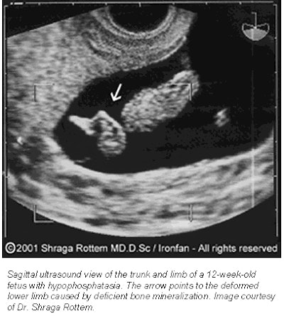 |
Adding to his case, early sonograms can detect a myriad of fetal problems. Of the several hundred types of anomalies detected by IRONFAN participants, 64 are cardiovascular and thoracic, 54 are musculoskeletal, 53 are gastrointestinal, 47 are found in the central nervous system, 23 are facial and nuchal defects, and 30 types are genitourinary, Rottem reports.
And the list is only expected to grow. In about one year, Rottem hopes to make the IRONFAN database accessible to everyone on the Internet. Theoretically, practitioners could compare their own data concerning suspected anomalies with the registry’s statistics and case histories.
Additional early pregnancy fetal anomaly information could be obtained by combining fetal mapping scans with existing screening tests. Consider the nuchal translucency test, which is a sonogram at 10 to 12 weeks used to screen for Down Syndrome and heart problems. This test is focused on the back of the fetus’ neck and doesn’t consider additional potential anomalies.
But according to the IRONFAN database, there are 115 types of fetal structural problems that may be detected in the 12-week nuchal scan. For this reason, Rottem said he believes women who receive the nuchal scan should also receive a more comprehensive, fully reimbursed fetal mapping scan, called Fetal SonoMapping, which can detect a wide range of problems. Merging these two 12-week tests -- nuchal scan for Down Syndrome screening and Fetal SonoMapping for the early detection of gross anomalies -- will result in a much more efficient use of resources, he said.
Ultimately, Rottem hopes that obstetric and radiology centers that scan at-risk patients will soon adopt fetal mapping scans. And, as the database grows and becomes easily accessible on the Internet, practitioners at major medical centers will realize the importance of time-oriented scanning, he said.
"Our hope is that professionals scanning at 18 to 20 weeks also recognize the importance of first-trimester scanning and time-oriented Fetal SonoMapping" Rottem said.
AuntMinnie.com contributing writer
June 12, 2001
Related Reading
MRI second-guesses fetal ultrasound, April 25, 2001
Click here to post your comments about this story. Please include the headline of the article in your message.
Copyright © 2001 AuntMinnie.com


