(Ultrasound Review) According to researchers from the Yeungnam University in Korea, ultrasound may be useful as a screening examination for patients suspected of having posterior cruciate ligament (PCL) injury.
They imaged 65 knees, 30 asymptomatic volunteers, and 35 patients with clinically suspected acute PCL injuries. Their findings were published in the May issue of Radiology.
Accurate clinical diagnosis of PCL injuries may be compromised by hemarthrosis, pain, muscle spasm, and swelling. The commonly performed posterior drawer test for assessing PCL injuries may be falsely negative due to limited knee movement, intact arcuate complex, or intact combined posterolateral corner ligament.
Originating from the lateral surface of the medial femoral condyle, the PCL runs in an oblique-sagittal course to insert onto the intercondylar area of the proximal tibia, 1cm below the articular surface.
For the ultrasound examination, patients were placed prone with the knee in a comfortable neutral position. Using a 5–10 MHz linear array, they scanned the distal half of the PCL in the longitudinal plane only. An assessment was made of the anterior-posterior (AP) thickness at the level of the tibial spine, the echogenicity, and the clarity of the posterior margin.
In order to avoid anisotropy artifact (falsely hypoechoic region due to nonperpendicluar scanning), pressure was exerted on the proximal end of the transducer to ensure perpendicular reflection of ultrasound.
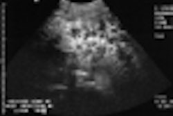
Scanning revealed that the normal PCLs were homogeneously hypoechoic, with a well-defined posterior border. Torn PCLs were heterogeneously hypoechoic with an indistinct posterior margin. There was a significant difference in the thickness of the torn PCLs compared with the normal PCLs. An AP thickness greater than 10mm was indicative of an acutely torn PCL.
The authors described arthroscopy as an invasive test that required multiple portals for viewing the entire length of the PCL and MRI as expensive.
"An acutely torn PCL thickens (>10mm), loses its sharply defined posterior border, and has a heterogeneously hypoechoic appearance. Ultrasound may be useful as a screening examination for patients suspected of having PCL injury and for deciding whether to perform more expensive MR imaging or surgical intervention," the authors concluded.
"Normal and acutely torn posterior cruciate ligament of the knee at US evaluation: preliminary experience"Kil-Ho Cho et al
Dept. of Diagnostic Radiology, Yeungnam University Medical Center, College of Medicine, 317-1 Daemyung-Dong, Nam-Ku, Taegu 705-717, Korea
Radiology; May 2001; 219: 375–380
By Ultrasound Review
June 20, 2001
Click here to post your comments about this story. Please include the headline of the article in your message.
Copyright © 2001 AuntMinnie.com


