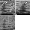
(Ultrasound Review) Ultrasound imaging plays an important role in the early identification of women at increased risk of fetal chomosomal abnormalities, through nuchal translucency measurement. Obstetricians from the universities of Varese and Bari in Italy, recently investigated whether there was a relationship between umbilical cord diameter at 10-14 weeks of gestation and chromosomally abnormal fetuses.Ultrasound in Obstetrics and Gynecology published their research in the March issue.
A study cohort of 784 consecutive patients was assembled. Chromosomal abnormalities were present in 17 fetuses. "The fetal karyotype was established by chorionic villus sampling or amniocentesis or at delivery in newborns with features suggestive of chromosomal abnormalities," they said.
All of the fetuses scanned had a crown-rump-length of 45-85 mm. The outer-to-outer cord diameter was measured twice and an average was calculated. Measurements were performed using a free-loop of cord in the longitudinal plane.
Mean cord diameter ranged from 3.2 mm at 10 weeks gestational age to 4.3 mm at 14 weeks. The authors have compiled a graph plotting cord diameter against gestational age showing the 5th, 50th and 95th percentiles. Comparing normal and abnormal fetuses, the cord diameter was above the 95th percentile in five of the abnormal fetuses (35.7%), and only 5% of normal fetuses. Combining nuchal translucency measurement and cord diameter, the combined rate of detection for fetal chromosomal abnormality was 85.7% compared with 71% for nuchal translucency measurement alone. The authors suggested some possible reasons for increased cord diameter including umbilical vein congestion and abnormal accumulation of fluid in Wharton’s jelly.
The authors hypothesized that "the underlying pathophysiologic mechanisms leading to an increase in umbilical cord diameter might be those that also explain the increased nuchal translucency in fetuses with abnormal karyotype, such as alterations of the extracellular matrix components of fetal venous congestion." They concluded that ultrasound measurement of the umbilical cord in the late first trimester appeared to identify a group of fetuses at increased risk of abnormal karyotype.
"First-trimester umbilical cord diameter: a novel marker of fetal aneuploidy"F Ghezzi et al
Dept of obstetrics and gynecology, University of Insubria, Varese, Italy
Ultrasound Obstet Gynecol 2002 May; 19:2335-239
By Ultrasound Review
May 29, 2002
Copyright © 2002 AuntMinnie.com
