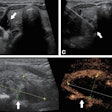
(Ultrasound Review) A prospective study reported in the October issue of the Journal of Ultrasound in Medicine details the use of sonographic markers using the Bayes theorem and likelihood ratios to determine the risk of Down syndrome.
Researchers at Harvard Medical School in Boston evaluated the significance of isolated and non-isolated sonographic markers in a large group of second trimester fetuses with Down syndrome, and compared this to a group of control fetuses.
The study was conducted over a period of 11 years and involved 164 fetuses with Down syndrome and 656 fetuses without aneuploidy. According to the authors, calculating likelihood ratios enables couples to "consider their individual risk estimate of carrying a fetus with Down syndrome and to decide whether to pursue amniocentesis, a procedure that carries a small but definite risk of pregnancy loss."
"We prospectively evaluated the mid-trimester sonographic features of fetuses with Down syndrome and compared them with euploid fetuses," they reported. Ultrasound findings evaluated included anatomical abnormality, increased nuchal fold (6 mm or greater), short long bones, pyelectasis (4 mm or greater), an echogenic intracardiac focus, and hyperechoic bowel.
Amniocentesis was performed on all fetuses at the time of ultrasound examination at 15-20 weeks gestational age. They reported "the presence of any marker resulted in sensitivity for the detection of Down syndrome of 80.5% with a false-positive rate of 12.4%. The absence of any markers conferred a likelihood ratio of 0.2, decreasing the risk of Down syndrome by 80%."
A thickened nuchal fold has an infinite likelihood ratio for Down syndrome, a short humerus had a likelihood ratio of 5.8, and structural anomalies carried a likelihood ratio of 3.3. Other markers had low likelihood ratios when found in isolation because of the higher prevalence in the unaffected population. "The likelihood ratios for the presence of 1, 2, and 3 of any of the markers were 1.9, 6.2, and 80, respectively," they discovered.
"Sonography cannot be used to diagnose or exclude aneuploidy,’ they said. "Although traditionally, a threshold of 1/270 has been used to offer invasive testing, the ultimate decision concerning prenatal diagnosis must be individual." Instead it provides a method of adjusting an existing risk for Down syndrome based of certain ultrasound markers.
According to the authors, "the potential of losing a pregnancy from a complication of amniocentesis is not equivalent to giving birth to a child with Down syndrome, and each outcome carries a different degree of devastation depending on the couple involved."
They concluded that " there are enough cumulative data to suggest that amniocentesis for advanced maternal age alone is no longer appropriate in the new millennium." They promote the use of first and second trimester ultrasound with biochemical analysis to predict individual risk assessment for Down syndrome before a decision is made regarding amniocentesis.
The genetic sonogram -- a method of risk assessment for Down syndrome in the second trimesterBromley, B et al
Departments of obstetrics and gynecology and radiology, Massachusetts General Hospital and Brigham and Women’s Hospital, Harvard Medical School, Boston
J Ultrasound Med 2002 October; 21:1087-1096
By Ultrasound Review
December 17, 2002
Copyright © 2002 AuntMinnie.com
