Three-dimensional ultrasound offers significant speed advantages over traditional 2D studies for transvaginal sonography, according to a pilot study published in the February issue of the Journal of Ultrasound in Medicine.
"Although standard 2D sonography is already quite efficiently used to perform a gynecologic transvaginal scan, 3D volume acquisitions can reduce scanning time by at least half, down to an average of one minute per scan," wrote the research team from Brigham and Women's Hospital in Boston. "The offline reconstruction of the volumes adds another 1.2 minutes per scan to the acquisition time; however, this still results in a total time savings, which is significant when 3D imaging is used compared with standard 2D transvaginal sonography" (JUM, February 2006, Vol. 25: 12, pp. 165-171).
The study team evaluated 35 consecutive patients who underwent gynecologic sonography. Following a standard 2D transvaginal scan that included measurements of the endometrium and abnormalities, four volume acquisitions were obtained. The volumes included a longitudinal acquisition of the uterus, as well as a transverse acquisition. The third and fourth acquisitions included both ovaries.
More than two weeks after the last examination in the study was completed, the volumes were then reviewed offline by the same sonologist who had obtained the original 3D acquisitions. The sonologist did not have access to any clinical information or any information from the original 2D exam at the time of review. Endometrium and other measurements were also performed on the volumes, according to the researchers.
After the review, the original sonography reports based on the 2D exams were obtained and compared with the information from the 3D volumes.
The 3D volume acquisitions took a mean time of 1.07 minutes (for a total of 37.54 minutes for all 35 cases), compared with a mean of 2.61 minutes for standard 2D scans.
Reconstruction, measurement, and interpretation of the volumes offline took 1.19 minutes for a total of 41.63 minutes for all 35 cases. In contrast, 2D required an exam time of 2.26 minutes. As a result, the 35 3D sonography cases were scanned and interpreted in 79.17 minutes, compared with a total of 91.46 minutes of 2D scan time (p = 0.47).
"Although the difference in time between the two methods for the performance and interpretation of the scan was only a total of 12 minutes for 35 patients, the difference in time spent obtaining the images in the ultrasound room was decreased by more than half when the 3D volumes were used, compared with the standard 2D technique," the authors wrote.
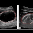
The researchers found no significant difference between the measurements of the endometrium, fibroids, and ovarian cysts when comparing 2D and reconstructed 3D images. There was also little difference in the ability of 2D and 3D sonography to identify the organs and abnormalities, according to the researchers.
"This study shows advantages of 3D over 2D sonography in the ability to obtain volumes rapidly, which can be reconstructed later, remotely and offline, and independently from the method of acquisition," the authors concluded. "This new capability is likely to become crucial for the future of sonography."
By Erik L. Ridley
AuntMinnie.com staff writer
February 8, 2006
Related Reading
Practical tips ease 3D/4D ultrasound, August 12, 2005
3D offers speed gains for fetal scanning, July 1, 2005
AMA says ultrasound in-utero 'portraits' are bad idea, June 22, 2005
4D ultrasound shows promise for improving fetal echocardiography, April 6, 2005
3D volume US shows promise for second-trimester fetal evaluation, March 17, 2005
Copyright © 2006 AuntMinnie.com


