When used interactively with a human reader, ultrasound computer-aided detection (CAD) technology can be a useful adjunct in the interpretation of breast ultrasound studies and can help prevent unnecessary breast biopsies, according to research from the University of Toronto.
In a retrospective study, a research team from the university's Joint Department of Medical Imaging found that the interactive use of ultrasound CAD yielded 98% sensitivity for malignant lesions, and could have avoided biopsy in 95 of the 102 benign lesions that had been biopsied. CAD did not perform as well, however, when used in an automated fashion.
"Using [ultrasound CAD] in an automated mode is not yet accurate enough for use in the daily interpretation of breast sonography," said Paula O'Donoghue, MD. "However, interactive [CAD] with a human observer allows for very accurate lesion assessment."
O'Donoghue presented the findings during a scientific session at the 2010 RSNA meeting in Chicago.
While ultrasound CAD is not used routinely today in breast sonography, the researchers believe that an accurate CAD system may open the door for the modality's use as a screening tool, O'Donoghue told AuntMinnie.com. Seeking to determine the diagnostic accuracy of an ultrasound CAD system based on American College of Radiology (ACR) BI-RADS lesion characterization, the researchers retrospectively reviewed 320 consecutive ultrasound-guided breast biopsy lesions from 2006.
The cases received at least two years of follow-up, and patients in the study had an age range of 16 to 92. The researchers included 266 lesions in the study, with 54 excluded due to file format reasons. Of the 266, 164 were breast carcinomas and 102 were benign breast lesions.
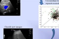
The blinded retrospective study evaluated the use of the B-CAD software (Medipattern, Toronto) with four expert readers. The B-CAD software performs lesion segmentation (i.e., finds the edge of the lesion) and then characterizes lesions using the BI-RADS lexicon. BI-RADS features are determined via separate algorithms that examine lesion edge, matrix, and the region surrounding the lesion.
The software generates a BI-RADS score and a corresponding editable "feature" report. It offers two BI-RADS choices: 2,3 (probably benign) or 4,5 (suspicious of malignancy) based on the lesion descriptive features, O'Donoghue said.
The researchers evaluated the CAD software in two modes: when used in an automated fashion and then in an interactive mode with a human reader.
In the automated mode, the CAD software processes images after the reader clicks on the center of the lesion. The software will automatically select what it determines to be the best fit for lesion segmentation, characterize the lesion using BI-RADS, and provide a BI-RADS score.
Just as in the automatic mode, the interactive mode begins by a reader clicking on the center of the lesion. CAD then processes the images. The algorithm provides six segmentation options for the lesion; once the reader selects the best choice, the CAD software completes processing and automatically characterizes the lesion, O'Donoghue said. A BI-RADS score is then provided.
The automated CAD yielded 62 (23%) false-positive cases that were segmented as BI-RADS 4,5. However, interactive CAD lowered the number of false-positive cases to seven (2.6%).
Interactive CAD also outperformed automated CAD in false-negative results, segmenting only two cases of malignancy as BI-RADS 2,3. The automated CAD system turned in 22 false negatives.
Of the 102 benign lesions in the study, use of interactive CAD could potentially have resulted in 95 fewer biopsies being performed, O'Donoghue said.
CAD performance by mode
|
The increase in accuracy was statistically significant (p = 0.00001), O'Donoghue said.
"These results obtained suggest that B-CAD imaging software, using the interactive segmentation mode, may be a promising addition to current clinical breast sonographic imaging," O'Donoghue told AuntMinnie.com.
Some improvement in lesion segmentation is required to increase the diagnostic accuracy of the automated B-CAD system, with the future potential for an ultrasound CAD application that can be applied to the real-time sonographic exam, she told AuntMinnie.com.
In addition, the potential yield of using B-CAD by readers with different expertise needs to be assessed prospectively, she said.
By Erik L. Ridley
AuntMinnie.com staff writer
February 15, 2011

