A large multicenter, international trial published in the February issue of Radiology has found that ultrasound with a shear-wave elastography protocol can significantly improve the specificity of breast ultrasound without hurting sensitivity.
In the 15-center prospective study, researchers determined that adding qualitative or quantitative features from shear-wave elastography images to B-mode ultrasound could reduce the number of unnecessary biopsies of low-suspicion BI-RADS category 4a masses (Radiology, February 2012, Vol. 262:2, pp. 435-449).
"It seems feasible based on our study that shear-wave elastography will help reduce unnecessary biopsies," said lead author Dr. Wendie Berg, PhD, of the University of Pittsburgh School of Medicine.
Low ultrasound specificity
While there is great interest in using screening ultrasound to augment mammography in women with dense breasts, the modality suffers from low specificity. In both the American College of Radiology Imaging Network (ACRIN) 6666 trial and in other studies of screening ultrasound, only 8% to 12% of masses that are biopsied only due to ultrasound findings have proved to be malignant, Berg said.
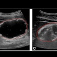
"We find many low-suspicion lesions on ultrasound that are mostly fibroadenomas and complicated cysts, which we know are very unlikely to be cancerous, but with standard techniques we cannot be sure without a biopsy," she told AuntMinnie.com.
Researchers and vendors have turned to elastography -- an ultrasound technique that measures the stiffness of tissue as an indicator of its malignancy -- as a possible solution. Berg and colleagues believed that adding tissue stiffness information from elastography could avert biopsy for most of the low-suspicion masses seen on ultrasound, even in the diagnostic setting, she said.
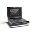
In the prospective study covering September 2008 to September 2010, 958 women with a median age of 50 received standard breast ultrasound using the scanner at their institution, followed by a quantitative shear-wave elastography exam using a prototype of the Aixplorer system that's commercially available from SuperSonic Imagine. Assessment of qualitative color shear-wave elastographic stiffness was performed independently.
Of the 939 masses that could be analyzed, 102 categorized as BI-RADS 2 were assumed to be benign. The reference standard was available for the remaining 837 lesions that were category 3 or higher. A BI-RADS categorization of 4a or higher was considered to be positive for malignancy. There were a total of 289 malignant masses in the study population, with a median mass size of 12 mm.
The researchers aimed to develop an algorithm that added one or more shear-wave elastographic features to B-mode ultrasound BI-RADS assessments. Focusing on the low-suspicion masses that cause the most treatment dilemmas, they emphasized the effect of adding elastographic features to the BI-RADS 3 or 4a lesion categories near the biopsy threshold level. Such features included oval shape, quantitative measures of elasticity, and visual assessment based on a colorized scale.
On its own, B-mode produced overall sensitivity of 97.2%, specificity of 61%, accuracy of 72.2%, and an area under the curve (AUC) of 0.950. Eight (2.6%) of the 303 masses categorized as BI-RADS category 3 were malignant, while 18 (9.3%) of 193 category 4a lesions and 41 (42%) of 97 category 4b lesions were malignant. Of the 57 category 4c lesions, 42 (74%) were malignant; 180 (96.3%) of 187 category 5 lesions were malignant.
All of the individual shear-wave elastography features were found to improve overall specificity in BI-RADS category 3 and 4a lesions without affecting sensitivity. But the researchers found that the best results were gleaned by adding visual color stiffness assessment (i.e., maximum qualitative elasticity) in the mass and surrounding tissue to the B-mode results.
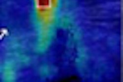
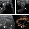
With maximum qualitative elasticity, a mass that was relatively soft and black or blue on the color overlay was considered to favor a benign etiology, while one that was red favored a malignant etiology; masses of green and yellow were considered to be indeterminate, according to the study team.
This visual color stiffness assessment correctly upgraded selected category 3 lesions and downgraded certain category 4a masses, resulting in an improved specificity from 61% (397 of 650) to 78.5% (510 of 650). The difference was statistically significant (p < 0.001). No significant impact on sensitivity was found.
In other findings involving individual elastography features, oval shape on shear-wave elastographic images produced specificity of 69.4% (451 of 650), while quantitative maximum elasticity of 80 kPa (5.2 m/sec) yielded specificity of 77.4% (503 of 650), both of which were statistically significant.
"We confirmed our impressions across a large 15-center international experience," Berg commented on the study results. "Even better, among the large number of masses that were felt to be probably benign, we expected that fewer than 2% would be malignant, but shear-wave elastography helped us identify some of those few cancers as well as to recognize with greater confidence that a low-suspicion mass was benign."
While the results show that shear-wave elastography may be able to reduce biopsies, it now must be proved in actual practice and decision-making, Berg said.
"Our study acquired the information and then, after the usual management, considered 'what if' possibilities," she said. "We need to see those borne out in real life."
Moderate- or high-suspicion masses should continue to be biopsied, even if they are "soft" on elastography, Berg noted. The researchers also hope that practitioners will not start biopsying stiff masses that would otherwise be recognized as typically benign.
|
Study disclosures The study was funded by SuperSonic Imagine. Among other disclosures, Berg reported that she has received consulting fees and payment for writing and reviewing the manuscript from SuperSonic Imagine. Several independent investigators listed as co-authors on the study also disclosed financial ties to the company related to the article, while several co-authors were SuperSonic Imagine employees. |

