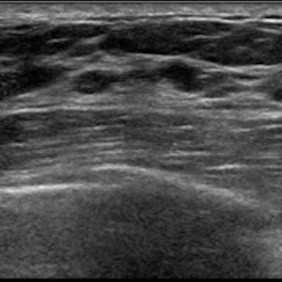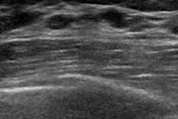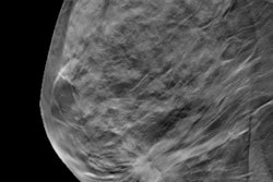
A diagnostic model that incorporates artificial intelligence (AI)-based quantitative analysis can significantly decrease false-positive rates for masses detected on screening breast ultrasound exams, potentially helping to reduce unnecessary biopsies, according to a study published online January 11 in Scientific Reports.
In a multicenter effort, researchers led by Dr. Soo-Yeon Kim, PhD, of Seoul National University Hospital in South Korea developed and tested a nomogram that encompasses radiologist assessments, as well as analysis of quantitative morphologic features extracted by a commercial deep learning-based computer-aided detection (CAD) software application.
They found that this diagnostic model could potentially cut the number of false positives from screening breast ultrasound by more than 40% without impacting sensitivity.
"[T]his is an effective method for reducing the false-positive biopsies associated with supplemental [ultrasound] exams," the authors wrote.
The researchers had sought to make use of AI technology for aiding the challenging task of characterizing breast masses spotted on screening ultrasound exams.
"Considering the small size and less obvious morphologic characteristics of breast masses detected using screening [ultrasound], the quantitative information derived from the [deep learning-based computer-aided diagnosis] software could provide additional details missed by the expert radiologists; furthermore, such information could prove to be substantially useful for the differentiation of breast masses while facilitating patient management in the clinical setting," they wrote.
The nomogram accounts for quantitative morphologic scores obtained prior to biopsy from S-Detect CAD software (Samsung Medison), along with patient age, mass size on ultrasound, and final BI-RADS assessment by the radiologist. The researchers developed and initially validated this model using 299 screening breast ultrasound exams that were retrospectively gathered from Severance Hospital and Samsung Medical Center in Seoul.
The nomogram's performance was then compared with that of final radiologist BI-RADS category assessments alone in an independent external test set of 164 women from Seoul National University Hospital.
| Nomogram vs. radiologists for characterizing masses detected on screening breast ultrasound | ||
| Radiologists' BI-RADS assessment ≥ 4A | Nomogram including AI analysis | |
| Sensitivity | 100% | 100% |
| False-positive rate | 97% | 45% |
| Biopsy rate | 98% | 48% |
| Area under the curve (AUC) | 0.51 | 0.87 |
With the exception of sensitivity (p = 0.317), the differences between the nomogram and the radiologists' BI-RADS assessment were statistically significant (p < 0.001).
"In comparison with the radiologists' BI-RADS final assessment alone, our nomogram incorporating the [AI software-] based quantitative information, patient age, and the BI-RADS assessments, improved AUCs and lowered the false-positive rates without decreasing the sensitivities in the differential diagnosis of breast masses detected by screening [ultrasound]," the authors wrote.
The researchers acknowledged that further improvements in performance must be demonstrated via larger and prospective studies before the method can be deployed in clinical practice. They also noted the current version of the S-Detect software does not automatically display these quantitative scores.
"In the future, providing the quantitative information as an output should be considered based on our results," the authors wrote. "The modified software would improve the diagnostic accuracy even in challenging situations, such as in the case of breast cancers detected using screening [ultrasound]."


