VIENNA - Multimodal tomography, a 3D nonionizing diagnostic imaging technology, may one day overtake mammography as the primary breast screening method considering its high sensitivity and specificity, according to a Thursday presentation at the European Congress of Radiology (ECR).
It's no secret that mammography is an imperfect modality -- it misses some cancers and doesn't work as well for certain women. However, it's cheaper than most other modalities and it's fairly easy to use, so mammography remains the primary method for breast cancer screening. That may change and multimodal tomography could take its place, according to Dr. Serafino Forte from the Institute of Radiology at University Hospital Basel in Switzerland.
"As predicted, multimodal tomography is a safe, comfortable, and specific technology, which may be a serious alternative to mammographic screening programs," said Forte, who presented research on behalf of Dr. George Zografos, PhD, from the University of Athens School of Medicine, and his team.
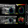
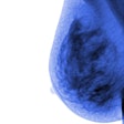
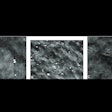
Multimodal tomography performs a 3D tomographic scan of the pendulant breast in a water bath using transmission ultrasound in a fixed-coordinate system. Multimodal tomography reconstructs multiple images for each coronal slice using measurements of refractivity and frequency-dependent attenuation and dispersion. The modality detects and differentiates lesions using fusion of the multimodal images. There is no need for compression and the patient lies prone for 12 minutes to scan one breast or 24 minutes to scan both.
The researchers found that multimodal tomography was able to detect and classify breast lesions down to 2 mm with 97.1% sensitivity and 98.5% specificity. Zografos and his team included 103 BI-RADS 4 female volunteers and compared histopathology results from biopsy of the suspicious lesions (size range, 2-28 mm).
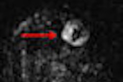
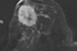
Multimodal tomography detected and classified 33 out of 34 malignant lesions in the biopsy samples. About half of the lesions (15) had a maximum dimension of 5 mm or less. The smallest detected lesion had a maximum dimension of 2 mm. Multimodal tomography had only one false positive in the biopsied lesions.
Limitations of the research included the selective patient sample and the high number of benign lesions (69).
"We in Basel believe in this new radiographic technology, and we are planning further studies in which we can compare multimodal tomography with traditional diagnostic tools like MRI and tomosynthesis, and of course histology," Forte said.
Neoadjuvant therapy and postproductive therapy will also be of interest, he said.


