Minnies winners, page 3
Scientific Paper of the Year
Winner: Detection of breast cancer with addition of annual screening ultrasound or a single screening MRI to mammography in women with elevated breast cancer risk. Berg WA et al, Journal of the American Medical Association, April 4, 2012. For AuntMinnie.com's coverage of this paper, click here.
The selection of this study as Scientific Paper of the Year highlights the interest in alternative imaging modalities as an adjunct to mammography for screening women with particular clinical conditions, in this case those with dense breast tissue.
Dense tissue is not only a recognized risk factor for cancer, it also tends to confound conventional x-ray-based mammography. Thus, researchers are trying to find other ways to improve upon mammography's sensitivity in these women.
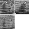
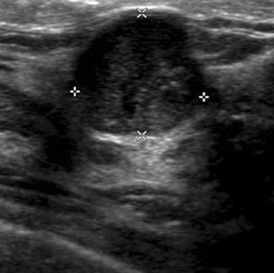
 Ultrasound of woman with heterogeneously dense breast tissue and negative mammogram. Scan demonstrates 1.5-cm grade III invasive ductal carcinoma, node negative. Image courtesy of Dr. Wendie Berg, PhD.
An ACRIN 6666 research team led by Dr. Wendie Berg, PhD, used MRI and ultrasound in conjunction with mammography in a population of more than 2,800 women with dense breast tissue who were seen across 21 institutions. Of these women, 612 received MRI scans.
A total of 111 breast cancers were found among 110 women, but mammography detected only half (59 cancers, or 53%) and had a sensitivity of only 52% on its own. Adding supplemental ultrasound uncovered an additional 5.3 cancers per 1,000 women in the first year, and 3.7 cases in the second and third years. Perhaps most significantly, in the women who received mammography, ultrasound, and MRI, use of the combined modalities produced a sensitivity of 100%, although specificity was 65%.
When analyzing the paper in an interview with AuntMinnie.com, Berg said that using all three modalities might be most appropriate in women with dense breast tissue and more than one risk factor for cancer. On the downside, false positives were an issue with the patient population, and many women simply denied a breast MRI scan, perhaps due to the requirement for contrast and issues with claustrophobia, Berg said.
Support for the study came from the U.S. National Cancer Institute and the Avon Foundation, providing funding through ACRIN.
Runner-up: Use of diagnostic imaging studies and associated radiation exposure for patients enrolled in large integrated health care systems, 1996-2010. Smith-Bindman R et al, Journal of the American Medical Association, June 13, 2012. For AuntMinnie.com's coverage of this paper, click here.
Best New Radiology Device
Winner: SonixEmbrace, Ultrasonix Medical
Products from major OEMs usually grab top honors in the Best New Radiology Device category, so it's notable that this year's winner -- SonixEmbrace from Ultrasonix Medical -- is a smaller company with a product that's not even on the market yet.
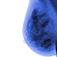
 SonixEmbrace automated breast ultrasound system. Image courtesy of Ultrasonix Medical.
Some of that probably has to do with the intense interest in finding alternative technologies to mammography, particularly for women with dense breast tissue. One of those technologies is ultrasound, and a growing number of clinical studies indicate that adding ultrasound to a conventional mammogram can improve the overall cancer detection rate.
SonixEmbrace is one of a new generation of automated breast ultrasound (ABUS) systems that are designed to overcome many of the limitations of freehand ultrasound when performed for breast imaging -- namely, that it's cumbersome to perform and is operator-dependent. These systems typically use an ultrasound transducer that automatically passes over the immobilized breast.
Patients lie face forward on the SonixEmbrace scanning bed while a rotating ultrasound transducer captures up to 800 ultrahigh-resolution slices in a 360° arc around the patient, according to the company.
Data from the scans are downloaded to the ultrasound platform in raw radiofrequency or B-mode format, and can be rendered with 3D viewing software or with a standalone desktop application. Data can also be integrated with CT or MRI scans, according to the company.
Ultrasonix Medical plans to begin the regulatory process for SonixEmbrace in 2013 with the FDA; a research version was introduced in April 2012.
Runner-up: Aquilion CXL CT, Toshiba America Medical Systems
Best New Radiology Software
Winner: iDose4 Premium Package, Philips Healthcare
With intense scrutiny being paid to CT radiation dose, it's no surprise that a software application that reduces dose was our expert panel's top pick for Best New Radiology Software.
The iDose4 Premium Package from Philips Healthcare is one of the recent generation of algorithms that use iterative reconstruction rather than traditional filtered back projection techniques to reconstruct CT data, generating higher-quality images at a sharply reduced dose.
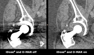 |
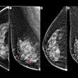
| Image courtesy of Philips Healthcare. |
Philips' Premium Package includes iDose4, the fourth generation of Philips' iDose iterative reconstruction technique, and technology to reduce artifacts from large metal implants. Called O-MAR (metal artifact reduction for orthopedic implants), the algorithm reduces artifacts that can result in a loss of anatomical information, impeding visualization of tissue and critical structures, according to the company.
The iDose4 Premium Package was launched in June at the International Society for Computed Tomography (ISCT) meeting in San Francisco. It is designed for the company's Brilliance CT 64-channel CT scanner and its Ingenuity and iCT families.
Runner-up: Syngo.plaza, Siemens Healthcare
Best New Radiology Vendor
Winner: CardioInsight
CardioInsight is developing ECVue, a 3D cardiac mapping system designed to generate real-time, whole-heart images of electrical activity by combining body-surface electrical data with 3D anatomical data obtained from CT scans. The firm was chosen by our expert panel as the Best New Radiology Vendor.
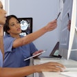
 Patients are scanned wearing ECVue vest. Image courtesy of CardioInsight.
The technology at the core of CardioInsight's electrocardiographic mapping (ECM) was developed at Case Western Reserve University by Dr. Yoram Rudy. The ECVue system gathers electrical information about the heart from a vest that is implanted with multisensory electrodes. The patient is scanned while wearing the vest, which gathers a view of the heart's electrical activity that can be combined with the anatomical information provided by CT.
CardioInsight believes the technology can offer an alternative to conventional catheter-based mapping methods. ECVue can help electrophysiologists better guide therapy, such as for localizing arrhythmia or optimizing the placement and setting of cardiac resynchronization therapy devices.
CardioInsight launched ECVue in Europe with its first installation in the U.K., and the firm now has additional installations in France and Germany. The company is working with the FDA on a regulatory submission for the technology, which may occur in late 2013.
Runners-up: Intrinsic Medical Imaging, Medspira, Scannerside
Click on the above links to learn more about each vendor.
Most Effective Public Service Campaign
Winner: Virtual Radiologic partnership with Doctors Without Borders/MSF
One of the more recent Minnies categories, the Most Effective Public Service Campaign, has proved popular as many in the radiology field turn their attention to ways in which they can use the power of medical imaging to improve the lives of those in underserved communities.
This year's winning initiative was announced in September 2011 as a partnership between teleradiology firm Virtual Radiologic (vRad) and international humanitarian organization Doctors Without Borders/Médecins Sans Frontières (MSF).
Through the partnership, vRad offers pro bono teleradiology consultations with its network of more than 400 radiologists to MSF physicians working at two locations, one in Nukus, Uzbekistan, and the other in Boguila, Central African Republic.
Uzbekistan is experiencing an outbreak of multidrug-resistant tuberculosis (MDR-TB), and MSF teams recently installed a digital imaging unit to help diagnose MDR-TB patients. vRad radiologists are also providing professional services to help with clinical consultations.
In the Central African Republic, MSF has installed a digital imaging system in Boguila. The country has been wracked by violence, and the healthcare system has completely collapsed; service provided by the collaboration between vRad and MSF is improving general medical care for both children and adults.
vRad has about 80 radiologists involved in the project, according to the company. Radiologists volunteer their time after their scheduled shifts, usually once a month, and the company has more radiologists interested in volunteering than it can accommodate. vRad also provides donated staff support for scheduling, implementation, and IT and operations center services.
vRad radiologists have also interpreted cases for several other MSF missions, including in Uganda and Malawi, and it also supports a non-MSF site in Haiti, Hospital Bernard Mevs. Overall, the company has read 500 studies so far across all volunteer sites.
Runner-up: Putting Patients First program, AHRA and Toshiba America Medical Systems
Previous page | 1 | 2 | 3

