Radiomic machine learning models could predict occult invasive cancer in women with ductal carcinoma in situ (DCIS), according to research published November 18 in Radiology.
The models, based on data from mammography features, achieved high marks in identifying candidates for active monitoring and suggesting women at high risk for upstaging who may need axillary lymph node sampling.
“Classification models for tumor upstaging may inform clinical management of patients with DCIS and mitigate the effects of under-sampling by core-needle biopsy,” wrote the researchers led by Rui Hou, PhD, from the Beijing University of Posts and Telecommunications in China and colleagues.
Many women with DCIS may not progress to invasive breast cancer, but surgery is recommended. Prior research suggests that about one in four women initially diagnosed with DCIS have their disease upstaged to invasive ductal carcinoma (due to under-sampling at core-needle biopsy).
Radiologists continue to explore the potential of radiomics in classifying diseases. Research has suggested that radiomic features of calcifications at mammography can predict occult invasive disease among women diagnosed with DCIS. However, the investigators noted that radiomics models need to be tested for their generalizability before being deployed into clinical practice.
Hou and colleagues evaluated the performance of radiomics models tested in three national datasets from the U.S., the U.K., and the Netherlands. The study included data collected between 2000 and 2021, and all women were diagnosed with DCIS at core-needle biopsy.
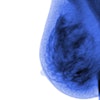
 Examples of correctly predicted lesions among three countries using the model trained on images from the U.S. dataset. Lesion annotations were provided by the study radiologists at each site. The regions of interest indicate lesion annotations.RSNA
The team developed its radiomic models by using 11 mammographic features and cross-validated logistic regression on each national dataset. It then performed round-robin testing on the other datasets.
The research included retrospective data from 1,498 women with an average age of 59 years (of these, 696 women were from the U.S., 618 from the U.K., and 184 from the Netherlands). Upstaging rates were 16.1%, 16.7%, and 14.1%, respectively.
On internal cross-validation, the models achieved the following areas under the receiver operating characteristic curve (AUCs): 0.675 (U.S. set), 0.603 (U.K. set), and 0.701 (Netherlands set). The model that was trained on the U.S. dataset achieved cross-national validation AUCs of 0.604 for the U.K. dataset and 0.682 for the Netherlands dataset.
Negative predictive values (NPVs) of the models ranged from 89% to 92%, while odds ratios ranged from 1.9 to 2.3, indicating that the models can help exclude cancers from active monitoring, the researchers wrote.
Finally, the models achieved positive predictive values of 30% and odds ratios ranging from 2.4 to 3.2, suggesting the models' potential to identify women at risk for upstaging and to help in surgical planning.
The study authors highlighted the promise these findings hold toward improving presurgical diagnosis of DCIS and management strategies.
“As we navigate the complex landscape of breast cancer diagnostics, our study offers a foundation for future research endeavors and clinical applications in the realm of radiomic feature analysis,” they wrote.
Read the entire study here.