Friday, December 4 | 11:50 a.m.-12:00 p.m. | SST01-09 | Room E450B
This Friday presentation will detail a sense-soothing imaging room that helps women relax while they undergo mammography, allowing better images to be acquired and possibly reducing false negatives.A study led by Shakira Sarquis-Kolber of Boca Raton Regional Hospital included 303 women who underwent mammography for two consecutive years. The women had one exam in a typical imaging room and another in a sensory-stimulating mammography room, which featured aromatherapy, videos with environmental themes, and relaxing sounds.
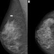
For each exam, the researchers calculated the amount of tissue imaged by measuring the posterior nipple line on the two mediolateral-oblique (MLO) images and the two craniocaudal (CC) images, as well as the compression force used.
The sensory-stimulating environment improved the amount of breast tissue captured in the mammography exam by 5% over the amount of tissue imaged in the typical exam room. There were no significant differences in the compression force needed to obtain this additional tissue, the researchers found.
The study suggests that incorporating sensory-stimulating features into the mammography room lessens women's anxiety and physical discomfort. It may also increase the sensitivity of the technology while reducing false negatives due to inadequate tissue visualization, Sarquis-Kolber and colleagues concluded.

