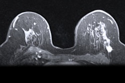
Ductal carcinoma in situ (DCIS) diagnosed with conventional breast imaging usually showed nonmass enhancement on breast MRI scans, with a larger median span for lesions than mammography and an increased cancer detection rate of 6.2%, in a study published August 3 in Radiology.
Researchers led by Dr. Shinn-Huey Chou from Massachusetts General Hospital also found that focal distribution of nonmass enhancement may indicate greater single wide-local excision success.
"Accurate preoperative prediction of DCIS biology and extent would assist with individualized risk assessment and treatment optimization," the study authors wrote.
DCIS makes up 20% to 25% of new breast cancer diagnoses in the U.S. While the 20-year breast cancer-specific mortality rate for women with DCIS is low at 3.3%, risk of breast cancer death remains 1.8 times higher than that of the general U.S. population.
Previous research has highlighted contrast-enhanced MRI's success in detecting DCIS with its high sensitivity and accuracy, but there are conflicting data on whether this leads to improved surgical outcomes, such as lower reexcision rates.
The investigators sought to assess breast MRI features of DCIS, its performance in identifying additional disease, and any associations of imaging features with outcomes.
The team looked at data taken from the Eastern Cooperative Oncology Group--American College of Radiology Imaging Network E4112 trial, including information from 339 women with biopsy-confirmed DCIS diagnosed with conventional imaging, including mammography with or without ultrasound.
The group found that the median preoperative span was larger on breast MRI compared with mammography, at 19 mm versus 12 mm (p < 0.001). MRI also led to additional biopsies in 19.5% of cases, generating a 32% positive predictive value, a 6.2% additional cancer detection rate, and a 14.2% false-positive rate.
The investigators also found that smaller MRI span and focal nonmass enhancement were linked to success of a single wide-local excision (p < 0.001).
Focal distribution was associated with greater single wide-local excision success compared with segmental or other distributions among women with DCIS manifesting as nonmass enhancement. However, this association was not significant in multivariable analysis adjusting for MRI size, background parenchymal enhancement, nuclear grade, and central necrosis at core needle biopsy among women with wide-local excision as their first surgery.
While basic DCIS features of nuclear grade and central necrosis were highly associated with DCIS score, the team found no evidence of qualitative imaging features that could predict DCIS score or upgrade to invasive disease at surgery.
"[Our findings support] a potential role of preoperative MRI in the prediction of DCIS at risk for requiring reexcisions and suggest bracketed localization approaches may be warranted in such lesions," the study authors concluded.


