Contrast-enhanced mammography (CEM) may be a reliable tool for assessing screening recalls, according to results published January 1 in the European Journal of Radiology.
Researchers led by Sara Marziali, MD, from the University of Milan in Italy, reported high diagnostic performance for CEM from at least three years of follow-up in women having their suspicious mammogram findings assessed.
“For these women at higher-than-average risk of breast cancer, CEM acted not only as a problem-solving tool but also showed an interesting screening performance in an intermediate-term timeframe,” Marziali and colleagues wrote.
CEM shows promise as a viable alternative to breast MRI for women who either have contraindications for MRI or lack access to MRI scanning.
Prior research suggests that CEM can help with inconclusive findings on screening mammography or ultrasound and preoperative assessments of disease extent in newly diagnosed breast cancers. The modality may also have utility in women with dense breasts and women at elevated risk, as well as for monitoring treatment response and post-treatment changes.
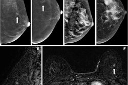
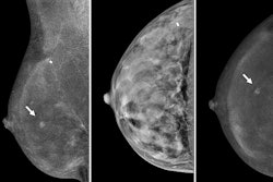
Marziali and colleagues studied CEM’s performance for assessing screening mammography recalls at three years of follow-up or more. Final analysis included 393 breasts from 198 women who had available biopsy or follow-up data.
The women were recalled after screening mammography at two Italian centers. They prospectively underwent CEM imaging between 2021 and 2024 alongside standard assessment through additional views, tomosynthesis, and/or ultrasound. The researchers independently evaluated both approaches, recommending biopsy or referral to a subsequent screening round.
Finally, the team calculated CEM’s per-diagnostic performance by taking histopathology and/or at least three-year follow-up data as the reference standard.
Of the total breasts included in the study, 316 (80.4%) were negative for disease either at the original biopsy or at follow-up. The researchers reported that 74 breasts had a malignant finding at the original biopsy, and three were interval cancers (0.8%) found during follow-up. The interval cancers included one case of ductal carcinoma in situ (DCIS) and two node-negative invasive cancers.
Per-breast analysis of the total breasts yielded the following results: 96.1% sensitivity, 94.9% specificity, 95.2% accuracy, 82.2% positive predictive value, and 99.0% negative predictive value.
The study authors highlighted that these findings “play in favor of CEM as a screening tool in women at higher-than-average risk.”
“As CEM represents a valid alternative to MRI, these models could be potentially applied to CEM as well,” they concluded.
Read the full study here.