Minnies finalists, page 3
Scientific Paper of the Year
Artificial intelligence versus clinicians: systematic review of design, reporting standards, and claims of deep learning studies, Nagendran M et al, BMJ, March 25, 2020. Learn more about this paper.

The first study up for the Scientific Paper of the Year title is on one of the hottest topics in radiology: artificial intelligence (AI). In the paper, a team of U.S. and U.K. researchers demonstrated that AI might not be all that it's cracked up to be.
Lead author Dr. Myura Nagendran of the Imperial College of London in the U.K. and colleagues reviewed studies that pitted deep-learning algorithms against expert clinicians for medical image interpretation tasks, including for radiology. Notable researchers on the project included Dr. John Ioannidis of Stanford University, Dr. Eric Topol of Scripps Research Translational Institute, and prominent AI researchers such as Dr. Hugh Harvey of Hardian Health in the U.K.
The review found major shortcomings of studies comparing the performance of AI and expert clinicians. Only 10 studies on the topic were the gold standard of randomized clinical trials, and of these, just two had been published.
The 81 nonrandomized trials in the review suffered flaws of their own, including that nearly three out of four papers had a high risk of bias. In addition, only 5% of studies gave full access to all datasets available, and just 7% gave full access to the code for preprocessing of data and modeling.
Based on the findings, the research team called for higher quality and more transparent evidence in order to avoid hype around AI and protect patients.
Radiology department preparedness for COVID-19: Radiology scientific expert review panel, Mossa-Basha M et al, Radiology, March 16, 2020. Learn more about this paper.

The second finalist for Scientific Paper of the Year was published just days before the first and is about the biggest story of the year, if not the decade: COVID-19. Written by the Radiology editorial board, the paper outlined best practices for radiology departments to address the novel coronavirus pandemic.
The board's initial guidance was both practical and remarkably farsighted for a time when the scientific community knew little about SARS-CoV-2 and its spread. Much of the guidance they published in March would soon be adopted throughout society, such as using screeners at hospital entrances and the radiology reception desk and requiring patients to wear masks during imaging and procedures.
Their advice also called for social distancing measures, including allowing staff to work from home, creating isolated onsite reading rooms, and using video conferencing for meetings -- although it's doubtful that even this team of authors could have predicted just how much time we'd spend on Zoom in 2020.
It's also easy to forgive the few best practices that missed the mark in retrospect. The one-month travel moratorium the group advised in March would have been greatly preferable to the widespread travel restrictions that will likely exist well into 2021. And had COVID-19 case numbers remained low in the U.S., perhaps it would have been sufficient for departments to follow the guidance to only quarantine staff with recent travel to countries with widespread, ongoing transmission.
Perhaps the paper's guidance was so pertinent because of the strength of the team behind it. Lead author Dr. Mahmud Mossa-Basha of the University of Washington in Seattle went on to become a prominent voice on the impact of the novel coronavirus on radiology. Other authors included Dr. Carolyn Meltzer of Emory University in Atlanta, a 2015 Minnies semifinalist for Most Effective Radiology Educator, and Dr. Danny Kim of NYU Langone Health in New York City, which faced its own surge of COVID-19 patients in April.
Best New Radiology Device
It's all about CT in the 2020 edition of the Best New Radiology Device competition in the Minnies. But this year's race offers two different takes on radiology's workhorse modality: one is a powerful high-end scanner optimized to perform cutting-edge imaging, while the other is a niche system dedicated to imaging a single organ.
Aquilion One / Prism edition CT scanner, Canon Medical Systems
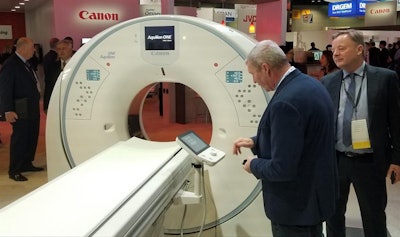 Aquilion One / Prism is Canon's new wide-area spectral CT scanner.
Aquilion One / Prism edition was designed by Canon Medical Systems to improve CT's ability to perform spectral imaging by combining artificial intelligence-based image reconstruction with Canon's wide-area detectors.
Spectral imaging involves the acquisition of images at two different energy levels. This enables users to analyze how different types of tissues -- such as malignant and benign structures -- respond to x-rays delivered at different energies. But spectral imaging has been somewhat slow to catch on in clinical practice.
Aquilion One / Prism edition is intended to revive spectral imaging by making it easier to perform in a clinical setting, without compromising on image quality, radiation dose, or workflow. In particular, Canon designed the scanner to address the workflow bottleneck that can throttle spectral imaging, by using deep-learning algorithms to improve how spectral results are integrated into the imaging chain, from acquisition through visualization. For example, in the PACS, spectral images are displayed next to conventional CT images.
For reconstructing images, Aquilion One / Prism uses Canon's Advanced Intelligent Clear IQ Engine (AiCE) algorithm, which distinguishes signal from noise to reconstruct sharper images at lower radiation dose. AiCE works on a variety of images, including brain, lung, cardiac, and musculoskeletal.
Canon launched Aquilion One / Prism at the RSNA 2019 conference; the scanner received U.S. Food and Drug Administration 510(k) clearance in February.
Somatom On.site portable head CT scanner, Siemens Healthineers
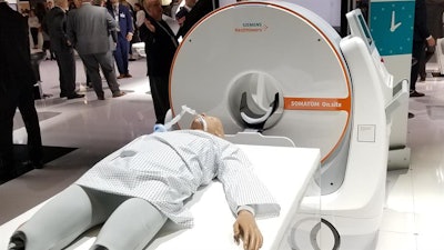 Somatom On.site is a new portable head CT scanner.
Also launched at RSNA 2019, Somatom On.site from Siemens Healthineers was designed with a specific purpose in mind: to take CT brain imaging to patients at their bedside in the intensive care unit (ICU).
Under the traditional model in radiology, patients are sent to the radiology department for scans, especially with a big-iron modality like CT. But On.site turns that model on its head, bringing the imaging to the patient.
On.site is mounted on a portable motorized trolley that can be moved with a handle; images are sent to PACS wirelessly. The system is a 32-slice scanner with a 0.7-mm detector width, and it acquires 2.4 cm of coverage per gantry rotation.
On.site sports an integrated drive camera that enables real-time viewing on the built-in touch display. Meanwhile, the scanner's telescopic gantry design allows the radiation source to move away from the patient during scanning, while the base and the front cover of the gantry remain stationary.
The scanner is also available with MyExam Companion, an intelligent user interface concept developed by Siemens that identifies the optimal acquisition and reconstruction parameters for patients using information such as their gender and age.
On.Site received 510(k) clearance from the U.S. Food and Drug Administration in July.
Best New Radiology Software
Clara COVID-19 AI Classification Model, Nvidia and the U.S. National Institutes of Health
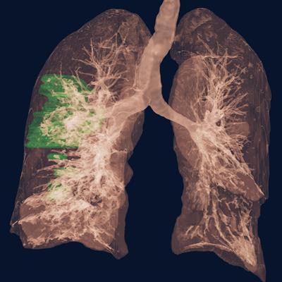 Clara COVID-19 AI Classification Model (for research use only). Image courtesy of Nvidia.
Nvidia and the U.S. National Institutes of Health (NIH) Clinical Center codeveloped the Clara COVID-19 AI Classification Model -- as well as a sister model for lung segmentation -- as part of a cooperative research and development agreement.
Using thousands of images that were labeled by radiologists at the NIH, the artificial intelligence (AI) models for chest CT exams were created in under three weeks using Nvidia's Clara application framework. Nvidia and the NIH then released the models for nondiagnostic use in May in hopes of facilitating the development of new AI algorithms by researchers around the world.
In research published in August, the classification model achieved an area under the curve of 0.949 for identifying COVID-19 pneumonia in testing on an independent dataset.
InferRead Lung CT.AI software, Infervision
InferRead Lung CT.AI, which received U.S. Food and Drug Administration 510(k) clearance in July, is an artificial intelligence-based application designed to assist radiologists in finding pulmonary nodules on chest CT scans.
After automatically performing lung segmentation, Lung CT.AI then identifies and labels nodules of different types for review by the radiologist, according to the company. Concurrent reading is supported.
Infervision believes that Lung CT.AI would be of particular value in CT lung cancer screening programs, helping to detect small lung nodules. As of July, the software had been deployed at over 380 hospitals and imaging centers around the world, and it was being used to process more than 55,000 cases each day, according to the company.
Best New Radiology Vendor
Hyperfine Research
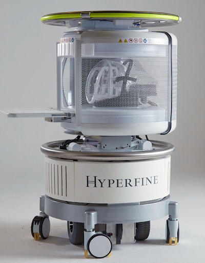 Hyperfine's point-of-care MRI scanner. Image courtesy of Hyperfine Research.
Hyperfine is bringing MRI to the point of care with its portable, low-field scanner, which mounts a 0.064-tesla magnet on wheels in a configuration that can be taken to the patients' bedside.
The scanner, which plugs into a standard electric wall outlet, uses ordinary permanent magnets that require no power or cooling to produce images. No special shielding is required.
The firm's initial version of the technology garnered U.S. Food and Drug Administration (FDA) clearance in February. The latest generation, Swoop, received FDA clearance in August and can be used for brain imaging in patients of all ages.
In the first clinical report of its performance, researchers recently reported that the scanner was feasible for neuroimaging patients in the intensive care unit.
RapidAI
Formerly known as iSchemaView, this artificial intelligence (AI) software developer for stroke imaging rebranded as RapidAI in February and has had a busy 2020.
The company landed U.S. Food and Drug Administration clearance for Rapid LVO, its software application for detecting large-vessel occlusions (LVO), as well as for its Rapid ASPECTS computer-aided diagnosis software, which is designed to automatically identify the Alberta Stroke Program Early CT Scoring (ASPECTS) regions of the brain on noncontrast CT and then generate an ASPECTS score to indicate early signs of brain infarction.
RapidAI also debuted RapidAI Insights, a data analytics platform that enables users to gain insight, standardize their stroke care processes, and optimize operational efficiencies, according to the vendor. In addition, the company acquired cerebral aneurysm management software developer EndoVantage.
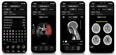 RapidAI's Rapid cerebrovascular imaging AI platform includes a range of stroke imaging and assessment applications. Images courtesy of RapidAI.
Additionally, RapidAI recently secured $25 million in funding to further develop its cerebrovascular imaging AI software around the world. In July, RapidAI announced that its Rapid cerebrovascular imaging AI platform has been used to analyze 1 million imaging studies from more than 1,600 hospitals in 50 countries.
Best Educational Mobile App
CTisus iQuiz, Dr. Elliot Fishman (iOS)
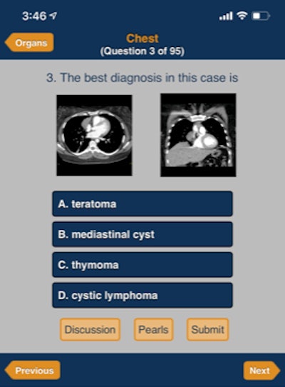 CTisus iQuiz. Image courtesy of Dr. Elliot Fishman
Dr. Elliot Fishman's CTisus family of mobile apps has produced winners of the Best Educational Mobile App award in the Minnies in three of the seven years the category has been offered. CTisus iPearls received the award in 2019 and 2017, while CTisus Critical Diagnostic Measurements in CT took the trophy in 2016.
This year's finalist, CTisus iQuiz, uses the quiz format to assist in the learning process for CT.
Now on version 11.5, the app features over 1,000 cases in 15 different categories that include case-specific "pearls" and an audio discussion of the case. Images, text, and sound are combined to enhance the learning experience.
As is the case for all CTisus apps, CTisus iQuiz is free to download and use.
Radiology Assistant 2.0, BestApps (iOS)
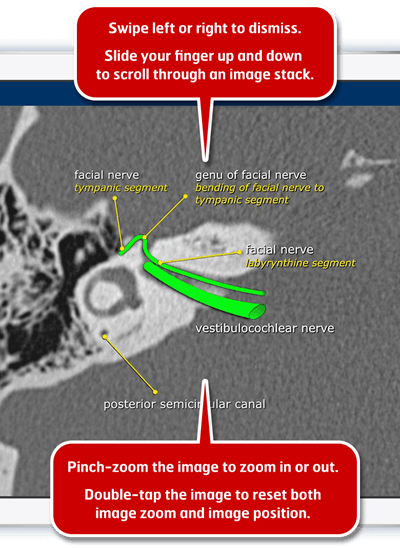 Radiology Assistant 2.0. Image courtesy of Dr. Wouter Veldhuis, PhD.
Radiology Assistant 2.0 has attracted much attention from the Minnies judging panel in recent years. This educational app developed by two Dutch radiologists is now a four-time finalist for Best Educational Mobile App, and it was also the winner in 2018. What's more, it's predecessor, Radiology Assistant, was a finalist in 2013.
Featuring peer-reviewed articles written by expert radiologists, Radiology Assistant 2.0 was developed by Dr. Wouter Veldhuis, PhD, of the University Medical Center Utrecht and Dr. Robin Smithuis of Rijnland Hospital Leiderdorp. Smithuis is the founder of the Radiology Assistant educational website and nonprofit organization.
The app, which is free to download but requires a subscription to access article categories, offers in-app purchases, supports full text searching in all articles, and allows users to view high-resolution versions of images in full-screen mode without leaving their place in the article, according to the developers.
Best Radiology Image
In this category, AuntMinnie.com members voted on our Facebook page on which of a set of 12 images should advance to the final round of Minnies voting. In the finals stage, winners will be selected by the Minnies expert panel.
The image below is derived from a study presented at the 2020 Society of Nuclear Medicine and Molecular Imaging (SNMMI) meeting. Researchers from Stanford University combined F-18 FDG PET with MRI to try to pinpoint the origins of chronic pain in patients and help their caregivers develop better management plans. Their findings indicate that PET/MRI combination could be an effective way for those who experience chronic pain to get relief.
![Adult male with decades of right neck pain, discomfort, and tightening following birth injury. The patient had failed multiple standard therapeutic maneuvers before presenting for F-18 FDG PET/MRI. Images show abnormally elevated FDG uptake (white arrows; SUVmax [maximum standard uptake value] = 1.2) observed in a linear pattern in the space in the posterolateral right neck, between the oblique capitis inferior and the semispinalis capitis muscles, where the greater occipital nerve resides. By comparison, the same region on the contralateral, asymptomatic side of the neck has an SUVmax = 0.7. This result encouraged a surgeon to explore the area. The surgeon ultimately found a collection of small arteries wrapped around the nerve in this location. The small arteries underwent lysis by the surgeon and the patient reported tremendous relief of symptoms. (A) Coronal thick slab MIP [maximum intensity projection] of F-18 FDG PET. (B) Axial LAVA [Dixon liver acquisition with volume acquisition] Flex MRI through the cervical spine. (C) Axial PET at the same slice as the axial MRI. (D) Fused axial PET/MRI. Image courtesy of Peter Cipriano of Stanford University; caption courtesy of the SNMMI.](https://img.auntminnie.com/files/base/smg/all/image/2020/09/am.2020_10_01_16_27_1916_2020_09_minnies_SNMMI_PET-MRI.png?auto=format%2Ccompress&fit=max&q=70&w=400) Adult male with decades of right neck pain, discomfort, and tightening following birth injury. The patient had failed multiple standard therapeutic maneuvers before presenting for F-18 FDG PET/MRI. Images show abnormally elevated FDG uptake (white arrows; SUVmax [maximum standard uptake value] = 1.2) observed in a linear pattern in the space in the posterolateral right neck, between the oblique capitis inferior and the semispinalis capitis muscles, where the greater occipital nerve resides. By comparison, the same region on the contralateral, asymptomatic side of the neck has an SUVmax = 0.7. This result encouraged a surgeon to explore the area. The surgeon ultimately found a collection of small arteries wrapped around the nerve in this location. The small arteries underwent lysis by the surgeon and the patient reported tremendous relief of symptoms. (A) Coronal thick slab MIP [maximum intensity projection] of F-18 FDG PET. (B) Axial LAVA [Dixon liver acquisition with volume acquisition] Flex MRI through the cervical spine. (C) Axial PET at the same slice as the axial MRI. (D) Fused axial PET/MRI. Image courtesy of Peter Cipriano of Stanford University; caption courtesy of the SNMMI.
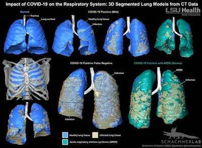
Adult male with decades of right neck pain, discomfort, and tightening following birth injury. The patient had failed multiple standard therapeutic maneuvers before presenting for F-18 FDG PET/MRI. Images show abnormally elevated FDG uptake (white arrows; SUVmax [maximum standard uptake value] = 1.2) observed in a linear pattern in the space in the posterolateral right neck, between the oblique capitis inferior and the semispinalis capitis muscles, where the greater occipital nerve resides. By comparison, the same region on the contralateral, asymptomatic side of the neck has an SUVmax = 0.7. This result encouraged a surgeon to explore the area. The surgeon ultimately found a collection of small arteries wrapped around the nerve in this location. The small arteries underwent lysis by the surgeon and the patient reported tremendous relief of symptoms. (A) Coronal thick slab MIP [maximum intensity projection] of F-18 FDG PET. (B) Axial LAVA [Dixon liver acquisition with volume acquisition] Flex MRI through the cervical spine. (C) Axial PET at the same slice as the axial MRI. (D) Fused axial PET/MRI. Image courtesy of Peter Cipriano of Stanford University; caption courtesy of the SNMMI.The second finalist in the Best Radiology Image category is an image from a case report of a patient with COVID-19 who was seen at a hospital in Louisiana. In findings published August 18 in BMJ Case Reports, researchers from Louisiana State University describe how they created 3D digital images that demonstrate the extent and distribution of COVID-19 disease in the respiratory system.
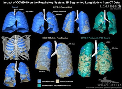 3D-segmented models of lung CT data show the distribution of COVID-19-related infection in the respiratory system. Image courtesy of LSU Health New Orleans.
Previous page | 1 | 2 | 3


