Dear AuntMinnie Member,
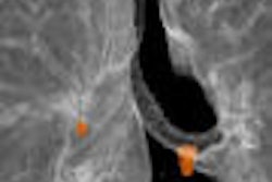
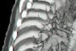
TORONTO - One of the most anticipated moments of the annual Society of Nuclear Medicine meeting is Dr. Henry Wagner's selection of the Image of the Year. This year was no exception, with Dr. Wagner bestowing honors on a 3D virtual bronchoscopy image produced by Stanford University researchers.
The image was generated by a PET/CT scanner using FDG as part of a study designed to validate a new acquisition and imaging protocol for producing 3D-rendered fusion images, according to staff writer Jonathan S. Batchelor, who was on hand to report on the story for our Molecular Imaging Digital Community.
The image displays FDG uptake in a primary cancer lesion and a mediastinal lymph node. Dr. Wagner described the image as emblematic of molecular imaging's unique ability to display structure, function, and biochemistry. See the image for yourself by clicking here.
We're featuring other articles from the SNM conference this week, including a story on the use of rubidium-82 PET for cardiac imaging to help heart patients avoid coronary angiography, and an article on using PET to track the effectiveness of psychotherapy for bulimia patients. You can read our coverage by visiting the Molecular Imaging Digital Community, at molecular.auntminnie.com.
Stay tuned for more stories from the SNM show in coming days, as well as reports in our Ultrasound Digital Community from the American Institute of Ultrasound in Medicine (AIUM) show in Orlando, and coverage in our Advanced Visualization Digital Community from the Computer Assisted Radiology and Surgery (CARS) meeting in Berlin.


