In the second part of our series on how to set up a 3D laboratory, AuntMinnie.com provides coverage of Dr. Eliot Siegel's suggestions on topics such as dealing with billing and coding issues and navigating the pitfalls of 3D labs. For part 1, click here.
The main revenue source to support 3D imaging is generated from reimbursement from Medicare and other health insurers. However, it's critical that services be billed only when appropriate, according to Dr. Eliot Siegel from the University of Maryland. 3D reconstructions must be performed on software that has the required clearances from the U.S. Food and Drug Administration (FDA) when billing Medicare.
 Dr. Eliot Siegel.
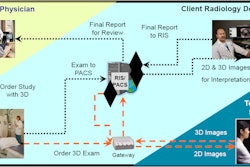
As an example of how 3D labs can be set up, Siegel described best practices in labs in operation at luminary sites. For example, the 3D lab at Stanford University utilizes an advanced visualization procedure manual that contains about 90 CT and MRI protocols, as well as quantitative data instructions. Referring clinicians can provide input to radiologists for consideration of additional protocols, Siegel said.
At the Massachusetts General Hospital (MGH) Radiology 3D Imaging Service, 3D imaging can be generated by referring physicians or radiologists, or by protocol, Siegel said.
A direct order from a referring physician for a specific scan with 3D imaging is considered to support the medical necessity of 3D imaging. In addition, medical necessity is often supported by the radiologist's documentation of patient history or exam indications, Siegel said.
Upon reading an examination, the radiologist can also request 3D studies if he or she notes positive findings and feels that additional views are necessary to address findings that affect the patient's well-being, Siegel said. Both the positive findings and the reason(s) for additional view(s) must appear in the radiology report to support the medical necessity of the 3D imaging.
Finally, 3D cases at MGH may also be performed and billed to Medicare via protocol if radiologists and/or referring physicians have determined that 3D reconstructions should be performed as part of the standard of care for a particular exam, he said.
If you don't have a 3D lab, billing for nonprotocol studies can be a real challenge, Siegel said. CT technologists typically don't have easy access to the ability to bill for studies, and radiologists typically don't have access at the workstation to direct entry of 3D billing information.
"Sometimes there can be difficulty or confusion about a radiologist designating a particular study to be billed subsequently, unless you have the workflow where, in the radiologist report or some other mechanism, the radiologist can let the billing office know that they dictated an extra paragraph or extra section about doing the 3D study," he said.
Coding
While the review of images in alternate display formats is already built into the CPT code definition for many procedures, separate coding for image reformatting is provided for selected procedures when such reformatting "entails complex rendering of the original image," Siegel said.
The 76376 and 76377 CPT codes should be reported in conjunction with the base imaging procedure code(s) when 3D rendering is performed in these procedures.
- CPT 76376 -- 3D rendering with interpretation and reporting of CT, MRI, ultrasound, or other tomographic modality, not requiring image postprocessing on an independent workstation
- CPT 76377 -- 3D rendering with interpretation and reporting of CT, MRI, ultrasound, or other tomographic modality, requiring image postprocessing on an independent workstation
According to the American College of Radiology (ACR) and the American Medical Association (AMA), both 3D codes require concurrent physician supervision of image postprocessing, 3D manipulation of the volumetric dataset, and image rendering, Siegel said.
For 3D reconstructions that don't require postprocessing on an independent workstation (such as those done on a CT console), the physician needs to discuss the need for 3D imaging with the technologist and supervise him or her in creating 3D images, Siegel said. In studies performed on an independent workstation, the physician is required to supervise and/or create the 3D reconstructions and adjust the projection to optimize visualization of anatomy and pathology.
"The 3D lab has to be under the careful supervision of the radiologist, but they don't talk about exactly how tight that process is," Siegel said.
The ACR and AMA state that the 3D rendering codes are intended specifically to address "complex renderings" such as shaded surface rendering, volumetric rendering, maximum intensity projections (MIPs), fusion of images from other modalities (but not PET/CT), and detailed quantitative analysis (segmental volumes and surgical planning), Siegel said.
The ACR has also emphasized that it's not appropriate to report the 3D rendering codes with certain selected procedures that already assume the performance of advanced visualization processing. In its CPT 2007 publication, the AMA provided a list of codes that should not be reported in conjunction with these codes.
In addition, 2D reconstructions are considered to be part of the base procedure code and should not be reported separately, Siegel said.
In other recommendations, 3D codes should not be used when 3D is not medically necessary, according to the ACR. When providing 3D rendering services, particularly in the outpatient setting, a "specific order is especially helpful to justify medical necessity," the organization noted.
The reformatting study should be documented in a separate report or in a separate section of the radiologist's report. When submitting for Medicare reimbursement, procedural CPT codes should be reported with diagnosis codes (ICD-9).
Variable reimbursement
Medicare coverage for image reformatting procedures varies. Medicare doesn't have a coverage policy that addresses the application of image reformatting in general, and coverage of these procedures is at the discretion of local Medicare contractors who process claims on behalf of the Medicare program, he said.
"Policy is highly variable," Siegel said. "So you have to talk to and negotiate with the local providers."
Private payors may rely on Medicare reimbursement policies, but others may not. Therefore, it's important to consult with individual private payors as well regarding coverage for this procedure, according to Siegel.
It's unclear how much these new 3D codes are currently being used by radiologists across the U.S. "There is tremendous variability in billing for advanced visualization," he said.
As for the question of whether an order is required by a referring clinician or if a radiologist can determine if advanced processing is indicated, the answer for hospital-based radiologists isn't 100% consistent, Siegel said. However, both the ACR and the AMA strongly recommend that a separate report be issued and that the medical necessity for the 3D rendering should be clearly stated.
For nonhospital-based imaging centers, a separate order from the referring physician is required. "You can't use the protocols that the hospital-based centers are using," he said.
3D codes cannot be used with studies where advanced visualization is already implied, including any CT angiography (CTA), MR angiography, nuclear medicine (including PET), CT colonography, and cardiac CT and CTA procedures.
If local expertise does not warrant doing billing onsite, it might be a good idea to consider outsourcing 3D billing to another 3D lab or service, according to Siegel.
Pitfalls
3D labs also have pitfalls to be aware of, including quality control (QC). QC is mandatory due to the wide variability in quantitative analysis tools and a lack of understanding of the limitations of the technology, Siegel said.
In addition, it would be a good idea to report results with a range in terms of level of confidence, rather than reporting specific numbers, he said.
Radiologists and clinicians have the potential to lose their skills in interacting with the dataset, he said. Similar to ultrasound, 3D image quality is also very dependent on technologist skill level.
Radiologists and technologists also need to be aware of some of the pitfalls of advanced visualization, Siegel said.
"Just because you have ability to render beautiful images does not mean they are optimally diagnostic and accurate," he said. "You need to have a lot of sophistication in setting this up."


