Tuesday, November 27 | 11:10 a.m.-11:20 a.m. | SSG05-05 | Room S405AB
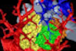
This scientific session will share the results of a multicenter study of a computer-aided detection (CAD) system for quantitative CT analysis of patients with idiopathic pulmonary fibrosis (IPF).CT is an essential imaging modality for detecting and diagnosing IPF; many studies have found a high correlation between the severity of CT findings, pulmonary function, and patient prognosis. Visual inspection and scoring are the conventional methods for quantitative evaluation of lung abnormalities, but visual scoring is a hard task for radiologists, said presenter Dr. Tae Iwasawa, PhD, of Kanagawa Cardiovascular and Respiratory Center in Yokohama, Japan.
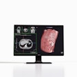
Visual assessment of a large number of CT image slices is also time-consuming and subject to substantial intra- and interreader variability. To provide automated and objective tools to assist with this problem, Kanagawa Cardiovascular and Respiratory Center and Yokohama National University have been developing a CAD system over the past 10 years for evaluating IPF, Iwasawa said.
A 2D version of the CAD system showed feasibility and utility in a previous single-center study, and the researchers now sought to conduct a multicenter, multivendor prospective study of the 3D version. In a test involving CT images of 80 patients with IPF obtained from four different types of CT scanners, CAD was determined to provide accurate and automatic assessment of pulmonary fibrosis, irrespective of CT scanner type, Iwasawa said.
"CAD-based volume measurements should be useful for detecting patients with rapid progression and, thus, help in the selection of aggressive treatment where possible," Iwasawa said. "Our goal is to provide the CAD system for everybody. We hope the system will be utilized more and more in clinical practice."


