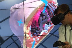
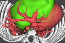
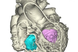
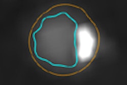
Researchers from the University of Minnesota are using 3D computer models generated from contrast-enhanced CT studies to create a digital anatomical library of human heart specimens.
In an online article published in the Journal of Visualized Experiments, a team led by Dr. Paul Iaizzo shared how surgeons and biomedical engineers at the university are combining computational technologies and CT scans of donated hearts to create a digital library of human heart specimens. The group believes this capability will assist in the design of cardiac devices.
Iaizzo's laboratory collects human heart specimens from organ donors that were not deemed viable for transplant. These organs can then be used for medical research and indexing.
Using contrast dyes and other reagents, the donated hearts are preserved and prepared to be in a diastolic state. This allows a deeper insight into the physiological attributes of the heart, including fluid capacity and pressure on the heart chambers, according to the researchers.
After the preserved hearts are scanned, computer models are generated to enable approximations and correlations to be established between various heart shapes and disorders, the researchers said.
