Dear Advanced Visualization Insider,
The development of new clinical applications for 3D printing is in full swing -- a clear boon for medical imaging. Among those applications is a project in Canada involving external-beam radiation therapy.
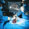
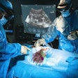
Doctors are taking data from CT scans to create 3D-printed pads, called boluses, for patients undergoing treatment after lumpectomy or mastectomy, among other applications. The customized 3D-printed boluses allow doctors to conform radiation dose to patient anatomy, keeping dose distribution even and effective. Find out how they're doing it in our Insider Exclusive.
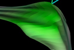
Farther west in Canada, another team is developing an algorithm for hip segmentation in infants using ultrasound. Investigators from the University of Alberta needed a fast and accurate segmentation scheme that could turn grainy, shadowy musculoskeletal ultrasound data into smooth and accurate 3D representations of the acetabulum for assessing developmental dysplasia of the hip, a condition that needs early treatment for optimal outcomes.
For all of its benefits in infant imaging, ultrasound comes with special challenges that are difficult to overcome, and an algorithm needs to skip past the acoustic shadows in the data to produce clean representations. See how the researchers pulled it off.
Breast CAD trouble
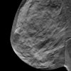
Meanwhile, trouble is brewing in breast imaging, where researchers have fought hard to earn a modest reimbursement for computer-aided detection (CAD) of mammography. The dark cloud appeared with the publication of a new study in JAMA Internal Medicine, in which radiologists obtained no better accuracy with breast CAD than without it -- contrary to the majority of literature on the topic. Could this development rock the industry and perhaps stymie the development of new technologies?
Also of concern -- the medicolegal kind -- is how radiologists deal with breast CAD markings. They typically discard them, but is that practice setting the stage for problems down the road? In an intriguing article about a talk from IT expert Dr. Eliot Siegel of the University of Maryland, experts question the common practice of discarding CAD markings after the exam.
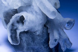
Finally, surgeons in Boston are using MRI to create 3D-printed heart models for surgical planning in young patients. The project aims to create a faster and more automated process to define the borders and contours of pediatric hearts and simplify the production of 3D models. Check out the models and learn more here.
For the rest of the news in high-tech imaging, we invite you to scroll through the links below, right here in your Advanced Visualization Community.
