Tuesday, November 29 | 9:50 a.m.-10:00 a.m. | RC303-06 | Room S504AB
Hoping to build a better interventional technique for doctors treating ventricular tachycardia with ablation guided by electroanatomic mapping (EAM), Italian researchers used CT to create a 3D heart model for each patient, overlaying EAM on the CT images to guide ablation. Hint: It works better than EAM alone."In patients suffering from ventricular tachycardia myocardial scars identified at late iodine enhancement, CT scans reflected with good accuracy ventricular tachycardia substrates identified through pathological voltages at EAM," Dr. Antonio Esposito, from Ospedale San Raffaele in Milan, explained to AuntMinnie.com.
The researchers sought to validate a CT acquisition protocol and image postprocessing for the creation of a 3D model of a heart containing information about cardiac anatomy and scarring. They compared the performance of ventricular tachycardia ablation with the model guided by EAM to EAM alone, which is the current treatment standard, Esposito said.
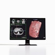
The study included 87 patients, 32 undergoing standard ablation and 55 undergoing dual-energy CT that included an angiographic scan and a delayed scan. These scans were used to create the 3D heart for each patient. The 3D models from CT were uploaded on standard Carto software and coregistered with the EAM data. The study explored procedure times, agreement between low EAM voltages and scar tissue, and success rates between the model heart technique and EAM alone.
"3D CT-derived heart models including information about cardiac chamber and scar can be merged to electroanatomic mapping systems in order to be used as real-time guidance to EAM and radiofrequency catheter ablation," Esposito said.


