Dear Advanced Visualization Insider,
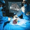
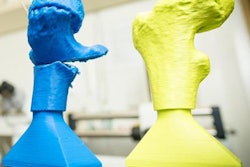
Mechanical thrombectomy -- an interventional stroke treatment that removes blood clots -- can be a challenging and stressful procedure for budding interventionists. Thanks to 3D printing, though, it's possible to practice and gain some experience before performing this vital treatment on live patients, according to a team from the University of Connecticut Health Center.
Using 3D printing technology along with MR angiography and CT angiography studies, a radiologist and a medical physics intern at the University of Connecticut created a brain perfusion phantom that enables physicians to practice threading a wire through the arteries that need to be accessed during a mechanical thrombectomy. 3D-printed models also offer potential in a number of other applications, including serving as a tool for understanding blood-flow dynamics and facilitating surgical planning, according to the group. You can read all about it in our Insider Exclusive.
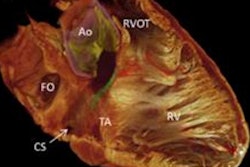
In other news this issue, a multinational research team has utilized 3D reconstruction of micro-CT images to visualize -- for the first time in 3D -- the group of cells that cause the heart to beat. The researchers applied volume rendering and segmentation techniques to create 3D representations of the cardiac conduction system on an intact human heart. How will this affect the understanding and treatment of diseased and congenitally malformed hearts? Click here to find out.
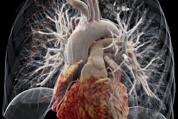
Cinematic rendering of medical images has drawn attention for its ability to create stunningly lifelike images from scans. In a pair of recent research papers, two separate groups shared their experience with the investigational technology and highlighted its potential benefits for complex studies and even for helping radiologists to create bonds with patients. But what exactly is cinematic rendering and will it offer an improvement over 2D images and 3D volume rendering? Click here to learn more.
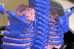
A pediatric radiologist at the University of California, San Francisco has developed imaging augmented reality software to aid in planning liver transplants and surgery. The software, which works with Microsoft's HoloLens virtual reality headgear to display CT images of the liver overlaid on a real-world background, shows promise for reducing surgical times and boosting surgeons' confidence in their treatment plans. Click here for all of the details.
We invite you to scroll through the links below for the rest of the news in advanced visualization and 3D printing, all here in your Advanced Visualization Community.


