Wednesday, November 28 | 12:45 p.m.-1:15 p.m. | IN228-SD-WEB4 | Lakeside, IN Community, Station 4
Using patient-specific 3D-printed models to plan for open surgical repair of the aorta may boost the procedure's accuracy and shorten operating time, according to this presentation being delivered on Wednesday.Repairing a tear in the thoracoabdominal aorta (TAA) is a complex and high-risk procedure requiring the reconstruction of vascular tissue. Though commercially available artificial grafts are available for the surgery, they do not take into account inherent variations in patient anatomy, noted Namkug Kim, PhD, and colleagues from the University of Ulsan College of Medicine in South Korea.
To meet this need, the researchers created 3D-printed aortas from the CT angiography scans of four patients with an aortic tear. They used the 3D-printed models as guides for constructing grafts to replace the damaged part of the aorta.
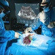
Kim and colleagues were able to measure the precise direction and location of surrounding arteries that would support the graft, allowing them to design a highly accurate graft and plan exactly where to situate it during surgery. These artificial grafts were individually tailored for each patient, and surgeons used them during open surgical repair of the aorta without the occurrence of any serious complications.
On average, surgery that relied on presurgical planning with the 3D-printed aortas took slightly less than eight hours -- considerably shortening the 12-hour operating time needed for more conventional techniques.
"This approach provides an individualized approach in open surgical repair of TAA based on the preoperative images, especially useful in anatomically challenging cases," the researchers concluded.


