Tuesday, November 28 | 3:30 p.m.-3:40 p.m. | SSJ20-04 | Room N229
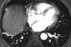
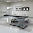
Boston researchers have developed a 3D printing technique for creating "biomimicking" phantoms that allow for the simulation of CT- and MR-guided thermal ablation.Image-guided interventions now account for a large percentage of all procedures performed, Dimitris Mitsouras, PhD, told AuntMinnie.com. There is a sharp learning curve to gain the necessary experience to perform these procedures well, and more challenging targets also are being explored, such as areas near the head, neck, and spine.
"However, there are very few simulation tools or phantoms that are currently available for CT- or MRI-guided intervention training," said Mitsouras, an assistant professor of radiology at Harvard Medical School and director of the Applied Imaging Science Laboratory at Brigham and Women's Hospital.
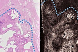
Mitsouras and colleagues eventually landed on 3D printing as a means to produce biomimetic phantoms with patient-specific anatomy that would facilitate the simulation of CT- and MR-guided procedures, namely thermal ablation.
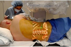
"In patient care, our 3D-printed biomimetic phantoms can already be used to optimize the safety and efficacy of ablation procedures," he said. "It allows the radiologist to try different arrangements of multiple probes and determine if one arrangement succeeds in sculpting the ablation zone so as to capture the entirety of a tumor."
These radiologically biomimetic phantoms have now assisted in the successful cryoablation of spine osteoid osteomas in two patients without leaving any residual tumor or causing any neural damage, according to Mitsouras.
"We hope this technology will begin to pave the way for interventional radiologists to be able to practice and simulate challenging procedures for specific patients, to assess the safety and accuracy of their treatment plan, and, importantly, to provide hands-on training for residents," he said.


