Sunday, November 29 | 11:25 a.m.-11:35 a.m. | SSA14-05 | Room E450A
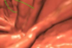
Volume-rendered 3D CT can provide time savings for detecting dislocated peroneal tendons, according to this scientific paper from researchers at the University of Iowa in Iowa City.CT studies are commonly performed for evaluating calcaneal fractures. When radiologists review the multiplanar reformatted (MPR) images of these CT studies on a 3D workstation, they can also access 3D volume-rendered images of the tendons, said Dr. Kenjirou Ohashi, a clinical professor of radiology at the University of Iowa.
"We found the 3D color images of tendons are easy to diagnose peroneal tendon dislocations, which are commonly associated with calcaneal fractures," he said.
Following up on a smaller study that found time savings for 3D volume-rendered images over MPR studies in this application (without a difference in diagnostic accuracy), the research team sought to compare the techniques in a larger study using 121 CT studies and three readers.
For the purposes of the study, consensus review of the MPR CT images by two experienced musculoskeletal radiologists served as the reference standard. Of the 121 studies, 43 were found to have peroneal dislocated tendons.
Three other musculoskeletal radiologists, with one to four years of experience with volume-rendered 3D images, blindly reviewed the volume-rendered images alone on a workstation. This approach led to sensitivities of 92%, 88%, and 81%, respectively, for the three readers.
"We believe this technique will save our time without compromising the diagnostic accuracy," Ohashi told AuntMinnie.com.


