Tuesday, December 1 | 11:30 a.m.-11:40 a.m. | SSG17-07 | Room S403A
In this Tuesday morning session, a U.S. National Institutes of Health (NIH) group will describe how computer-assisted image analysis may facilitate classification and guide clinical management of renal neoplasms.The NIH researchers wanted to improve upon the use of 2D measurements for renal cancer diagnosis, which can be time-consuming and highly variable. They sought to investigate the potential of computer-aided analysis of contrast-enhanced abdominal CT data for quantification and analysis of kidney lesions.
The software application supports 3D analysis and allows for the serial monitoring of lesions, according to presenter Marius George Linguraru, Ph.D., of the NIH's department of radiology and imaging sciences in Bethesda, MD.
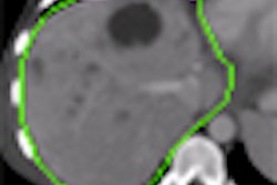
The group utilized the application to segment 116 renal lesions from 40 contrast-enhanced, three-phase abdominal CT studies. The tool not only accurately quantified cystic, solid, and mixed renal tumors, it also classified cancer types into four categories with statistical significance, according to the researchers.
"Computer-assisted image analysis of renal neoplasms on abdominal CT may facilitate their noninvasive classification and guide clinical management," Linguraru said. "The advance of strategies for cancer treatment and understanding causes of therapeutic failure will also rely on a combination of adequate perception of the underlying biological mechanisms and noninvasive imaging biomarkers."


