Friday, December 4 | 10:50 a.m.-11:00 a.m. | SST10-03 | Room S402AB
A computer-aided segmentation method can accurately provide volume and Response Evaluation Criteria in Solid Tumors (RECIST) measurements for head and neck tumors, according to researchers from Goethe University Frankfurt in Germany.Tumor volume has been shown to represent a prognostic factor in squamous cell carcinoma in the head and neck region, but its routine evaluation in staging CT exams has been hindered by the need for cumbersome manual slice segmentation, said presenter Dr. Ralf Bauer.
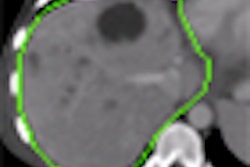
In their talk, the researchers will describe their evaluation of a semiautomated tumor volume assessment application (Siemens Healthcare, Erlangen, Germany) in 18 tumors from 15 patients. A radiologist first measured the RECIST diameter and volume of the tumors manually and then used the dedicated segmentation software tool. The manual slice segmentation by the radiologist served as the gold standard.
The researchers found that the software yielded highly accurate assessment of RECIST diameter and tumor volume (r = 0.99). The software also led to time savings of approximately 700%.
"This can be the first step of routinely assessing tumor volume and including the results in the radiological report," Bauer said.


