Tuesday, December 1 | 10:40 a.m.-10:50 a.m. | SSG06-02 | Room E353C
As ultrasound contrast becomes more widely used (at least outside the U.S.), software is becoming available to analyze contrast-enhanced ultrasound (CEUS) images. In this Tuesday morning presentation, Swiss researchers will discuss their use of a commercially available software application for analyzing CEUS images of focal liver lesions.Researchers from University Hospital in Lausanne wanted to examine the use of a contrast agent (SonoVue, Bracco Imaging, Milan, Italy) in conjunction with dedicated CEUS analysis software (SonoLiver, Bracco). SonoLiver analyzes dynamic vascular patterns in different liver phases, such as arterial, portal, and late phases, and includes both perfusion imaging and quantification tools for parametric image analysis.
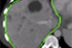
The researchers have further enhanced the software by adding color coding of different contrast enhancement patterns: yellow when the lesion remains more enhanced than the surrounding parenchyma, and green when it changes from hyper- to hypoechoic with regard to liver parenchyma.
Presenter Dr. Anass Anaye will discuss how the researchers used a population of 145 patients with 146 liver lesions, all of whom received a bolus injection of SonoVue and were imaged with nonlinear imaging modes at low mechanical index. The group used SonoLiver to calculate different liver perfusion parameters such as time to peak, rise time, wash-in rate, maximum intensity, and wash-out rate.
Readers used the software to analyze the wash-out phase of liver perfusion to make a diagnosis. Their performance was measured according to receiver operator characteristics (ROC) analysis, calculating sensitivity, specificity, and positive and negative predictive values. Final diagnoses were confirmed by MRI, CT, or biopsy.
Of the 146 lesions, 113 were malignant and 33 were benign. The readers using SonoLiver classified 110 lesions correctly for a sensitivity of 97.3%, with three false negatives. Of the benign lesions, 30 of 33 were correctly classified as negative, yielding specificity of 93.9%. The researchers said that there was a significant difference in contrast enhancement kinetic parameters between malignant and benign tumors.
"This study confirms that CEUS has a high specificity for the characterization of [focal liver lesions] and that a parametric image may adequately summarize the behavior of a [focal liver lesion] after the injection of the ultrasound contrast agent," according to co-author Dr. Jean-Yves Meuwly. "Quantitative data from the enhancement process also has a high diagnostic accuracy."


