Thursday, December 3 | 10:30 a.m.-10:40 a.m. | SSQ10-01 | Room S402AB
A team of Microsoft researchers will present a software algorithm that can automatically detect and localize anatomical structures in 3D CT images.The application of automatic recognition techniques in medical imaging could be useful for both radiologists and clinicians. For example, radiologists spend at lot of time looking at images, a task that could be made more efficient through the use of these methods, according to presenter Antonio Criminisi.
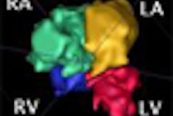
"If their computer was able to recognize [automatically] position, orientation, and extent of several anatomical structures within a scan, then the radiologist [could] click on the word 'mitral valve' to be taken to the most appropriate and useful view of that structure; i.e., the view which is most relevant for an accurate diagnosis," Criminisi told AuntMinnie.com. "This would greatly reduce wasted time and improve the radiologist's workflow."
Further applications of this technology include content-based image searching and initializing a more organ-specific type of image processing, Criminisi said.
The researchers trained and tested their technique using a database of labeled CT images. The algorithm turned in excellent accuracy in detecting organs such as liver, heart, kidneys, lungs, eyes, and head. In addition, it also yielded localization accuracy of around 0.5 cm for these organs, according to the researchers.


